
![]()
Echo basics: Valve Views
Overview
Heart valve disease affects approximately five million adults in the USA. It is a significant contributor to morbidity and mortality, particularly in aging populations.
Etiology and Classification
Valve dysfunction is broadly categorised into:
- Primary valve disease: Structural abnormalities of the valve itself.
- Secondary (functional) valve disease: The valve is structurally normal but its function is impaired due to abnormalities in surrounding structures, such as the myocardium or great vessels.
Types of Valve Dysfunction
Valve dysfunction manifests as:
- Stenosis: Narrowing of the valve orifice, impeding forward flow.
- Regurgitation (or incompetence): Incomplete valve closure causing backward flow of blood.
Causes of Valve Disease
- Congenital abnormalities
- Age-related calcification
- Inflammation (e.g., rheumatic heart disease)
- Infection (e.g., infective endocarditis)
Congenital and acquired valve abnormalities
Congenital Abnormalities
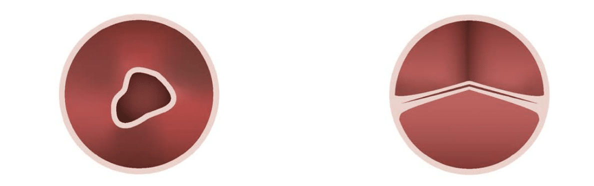
- Variations in cusp number are the most common congenital finding in adults on echocardiography, particularly affecting the aortic valve.
- Bicuspid aortic valve is the most frequent congenital anomaly, which predisposes to early calcification and dysfunction.
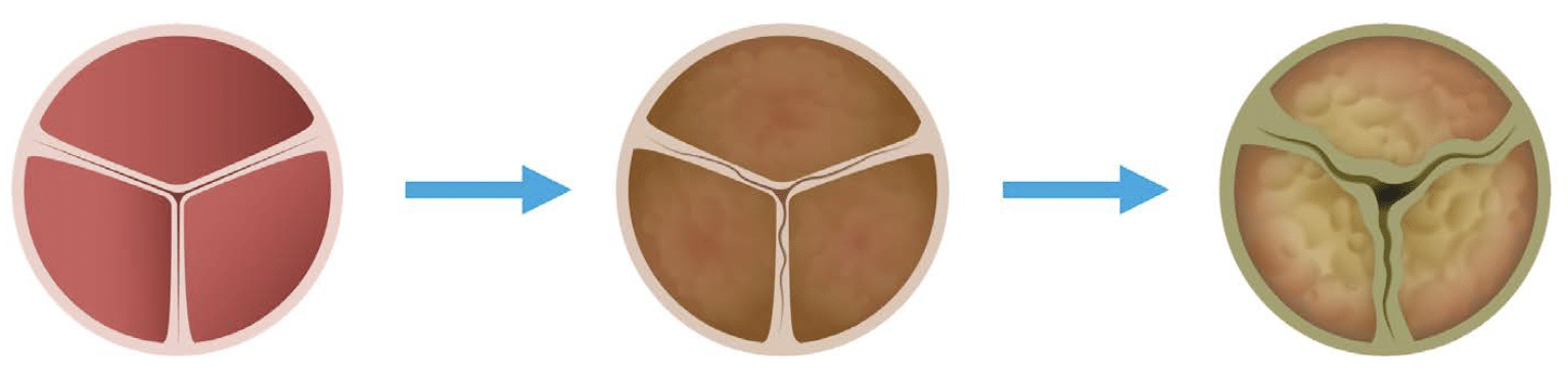
Calcific Valve Disease
- Calcific valve disease is increasingly common with population aging.
- There are potential links between calcific aortic valve disease and atherosclerosis, with shared risk factors and overlapping pathophysiological mechanisms.
Pathogenesis:
- Endothelial damage to valve tissue
- Lipid infiltration and inflammation
- Progressive calcification, leading to:
- Reduced valve flexibility
- Impaired valve leaflet movement
- Eventual stenosis
Endocarditis
- Endocarditis refers to inflammation of the endocardium, the inner lining of the heart and valves.
- It is usually due to infection and is considered rare but life-threatening.
- Common causative organisms include Staphylococcus aureus and Streptococci.
- Prompt diagnosis and treatment are essential due to high morbidity and mortality.
Rheumatic Valve Disease
- Rheumatic fever, a complication of untreated Group A Streptococcal infection, can result in rheumatic heart disease, primarily affecting the mitral and aortic valves.
- It is now rare in developed countries due to early antibiotic treatment.
- Remains prevalent in low- and middle-income countries, where it is a major cause of valvular heart disease in younger populations.
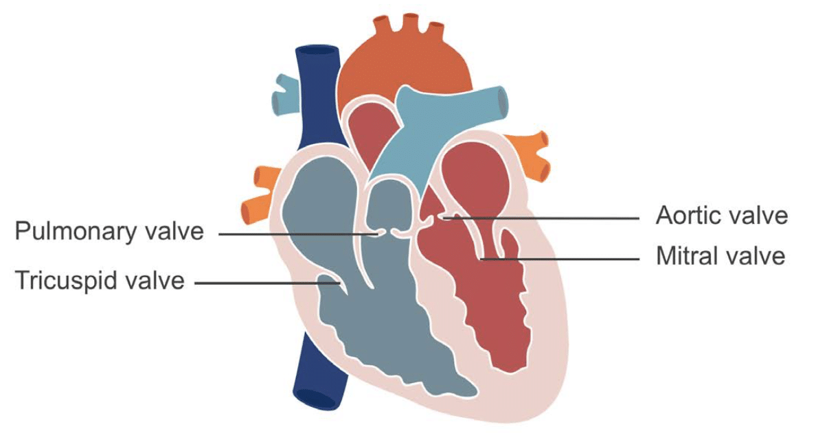
Viewing the valves with echocardiography
The valves can be seen from almost all the standard views of the heart but a full evaluation of the valves will require imaging from a variety of windows
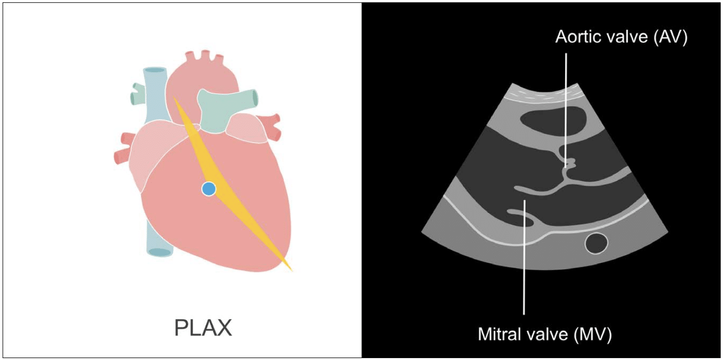
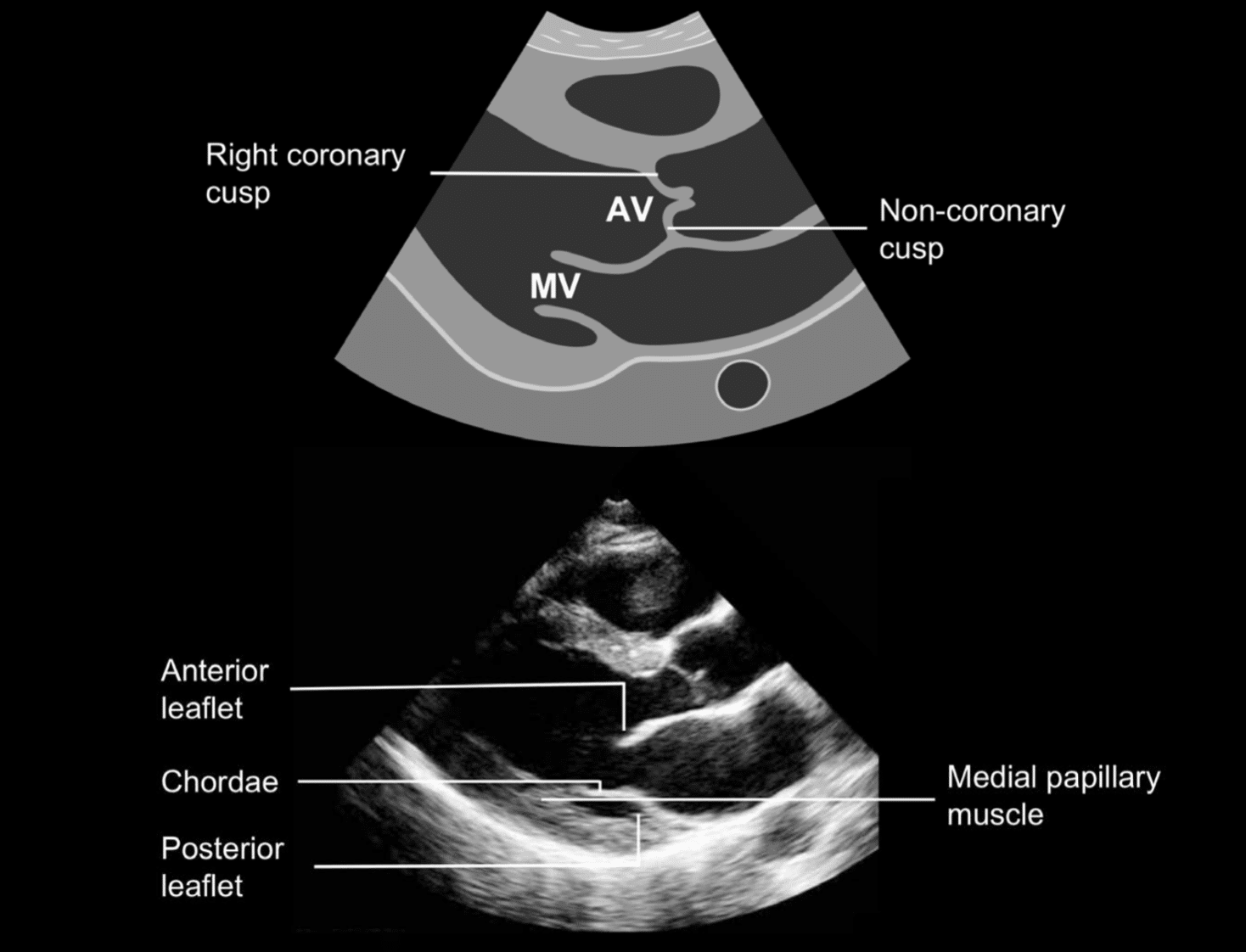
PLAX: Parasternal long-axis view
The standard parasternal long-axis approach usually provides clear views of both the aortic valve and mitral valve.
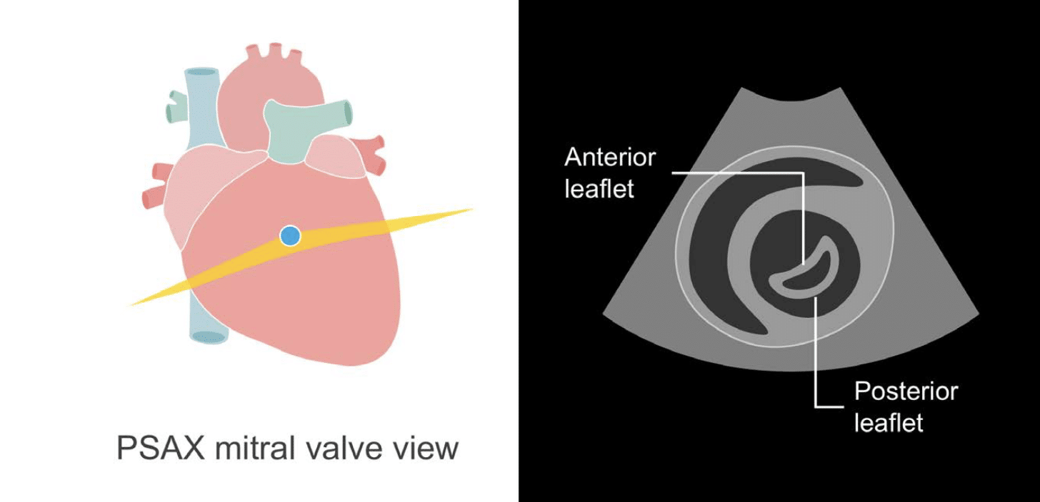
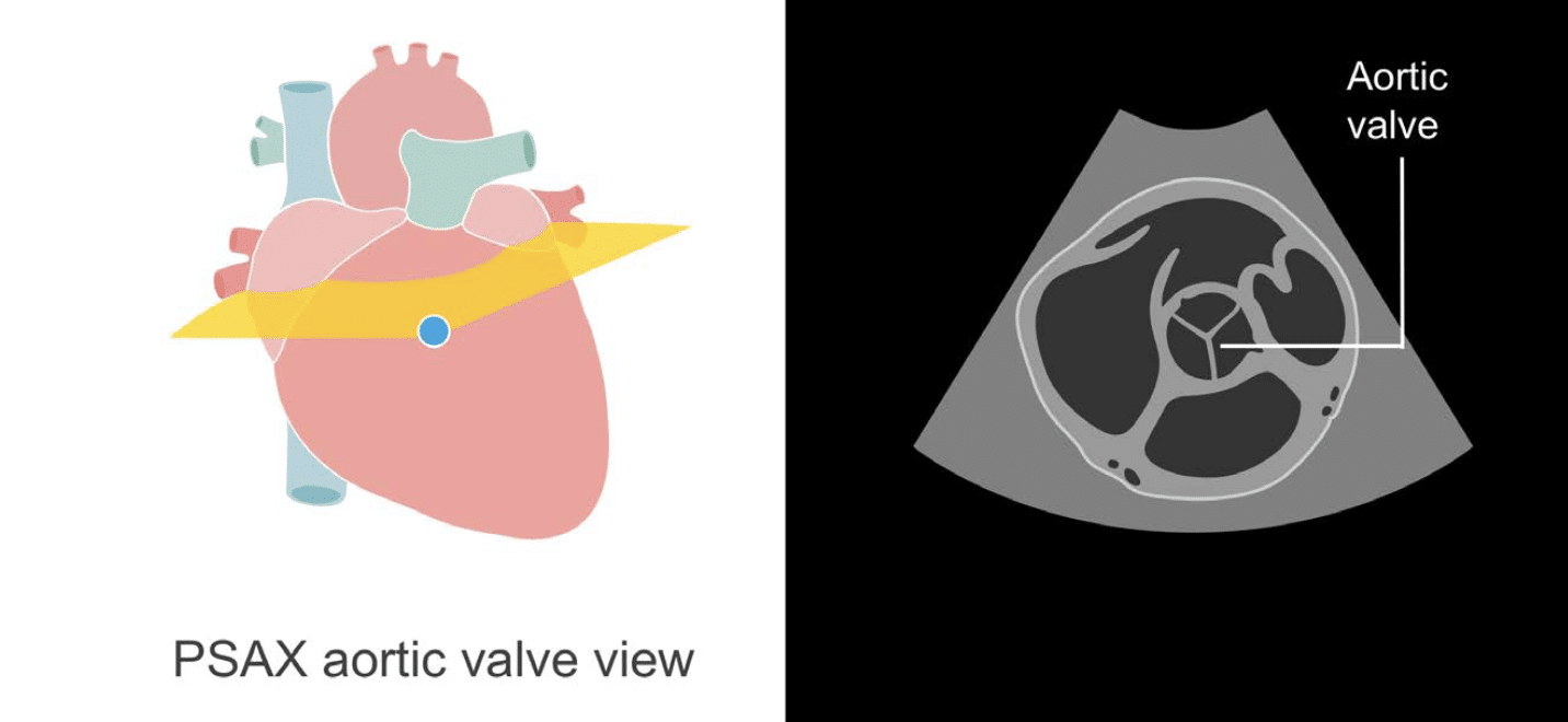
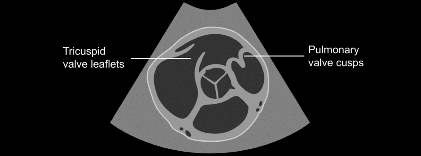
PSAX: Parasternal short-axis view
Obtained by rotating the probe 90 degrees in a clockwise direction from the long-axis view. This view gives useful structural information, particularly for the aortic valve and mitral valve. The pulmonary valve and tricuspid valve are also seen.
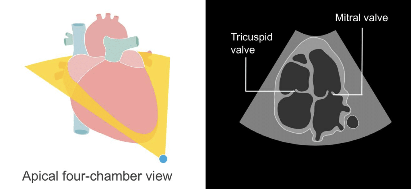
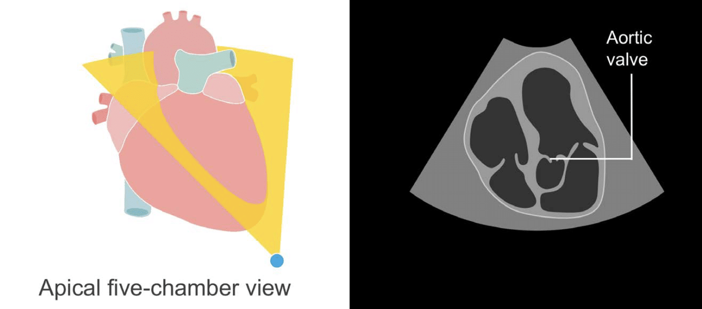
Apical views: 4-chamber and 5-chamber
The apical four-chamber view allows us to see the mitral valve and tricuspid valve and their associated structures. The so-called five-chamber view allows the aortic valve to be seen.
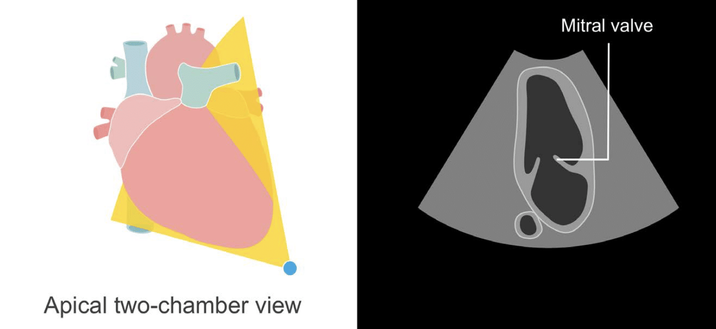
Apical views: 2-chamber and long axis
The apical two-chamber view shows the mitral valve, and the apical long-axis view shows the aortic valve and mitral valve.

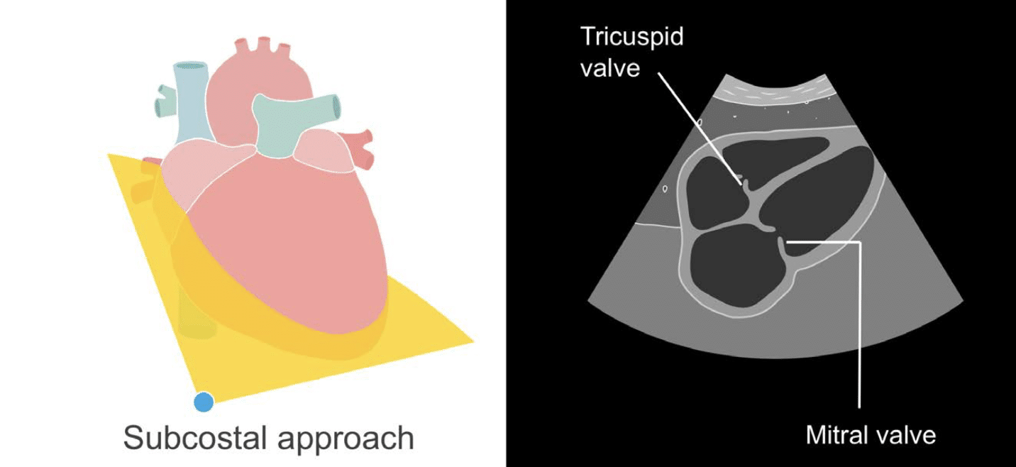
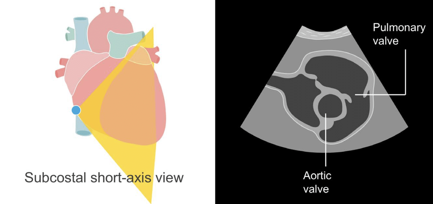
Subcostal views
This is an edited excerpt from the Medmastery course Echo Masterclass – The Valves by Chris Eggett, PhD. Acknowledgement and attribution to Medmastery for providing course transcripts.
Additional echocardiography resources:
- Na, M. Echo Masterclass: Left Ventricular Strain. Medmastery
- Monteiro, C. Echo Masterclass: The Right Heart. Medmastery
- West, C. Echo Masterclass: Adult Congenital Heart Disease. Medmastery
- Naderi, H. Echo Masterclass: The Power of 3D Imaging. Medmastery
Radiology Library: Echocardiography basics
- Eggett C. Echo basics: Valve Views. LITFL
- Eggett C. Echo basics: Valves, Measurements and Reports. LITFL
- Eggett C. Echo basics: Mitral valve. LITFL
- Eggett C. Echo basics: Mitral Regurgitation. LITFL
- Eggett C. Echo basics: Mitral Stenosis. LITFL
- Eggett C. Echo basics: Aortic Valve. LITFL
- Eggett C. Echo basics: Aortic Stenosis. LITFL
- Eggett C. Echo basics: Aortic Regurgitation. LITFL
- Eggett C. Echo basics: Tricuspid Valve. LITFL
- Eggett C. Echo basics: Pulmonary Valve. LITFL
- Eggett C. Echo basics: Prosthetic Valves. LITFL
Further reading
- Otto CM. Textbook of Clinical Echocardiography. Elsevier. 7e, 2023.
- Houghton AR. Making Sense of Echocardiography: A Hands-on Guide. 3e 2023
- Nishimura RA, Otto CM, Bonow RO, Carabello BA, Erwin JP 3rd, Guyton RA, O’Gara PT, Ruiz CE, Skubas NJ, Sorajja P, Sundt TM 3rd, Thomas JD; ACC/AHA Task Force Members. 2014 AHA/ACC Guideline for the Management of Patients With Valvular Heart Disease: a report of the American College of Cardiology/American Heart Association Task Force on Practice Guidelines. Circulation. 2014 Jun 10;129(23):e521-643
Echocardiography Essentials

Cardiac physiologist, echocardiographer, and Professor of Healthcare Science Education, Faculty of Medical Sciences at the University of Newcastle, UK. I direct post-grad programs at the Faculty of Medical Sciences, run an echo clinic at the Freeman Hospital, and teach transthoracic echocardiography to specialists in critical and emergency care and anaesthetic settings