


Gartland classification
The Gartland classification is a radiographic system for extension-type supracondylar fractures of the humerus in children. It grades injury according to displacement, preservation of cortical contact and fracture stability.
Type I fractures are undisplaced or minimally displaced; type II fractures are displaced but retain posterior cortical contact; type III fractures are completely displaced with no cortical contact; and type IV fractures are unstable in both flexion and extension because no functional periosteal hinge remains.
The classification provides a common language for emergency clinicians, radiologists and orthopaedic surgeons. Increasing grade generally reflects increasing instability and a greater likelihood of reduction and percutaneous fixation. Type I fractures are usually treated with immobilisation while type III and IV fractures generally require operative stabilisation. Management of type II injuries depends upon alignment, rotation, translation and the ability to maintain reduction.
Classification should not replace full clinical assessment. Neurovascular status, swelling, skin compromise, open injury, associated fractures, coronal alignment and medial-column collapse may alter management independently of the Gartland type. Type IV injuries are usually diagnosed dynamically during reduction under anaesthesia rather than from the initial static radiographs.
Modern use and terminology
The most consistently defined contemporary system is:
| Type | Definition |
|---|---|
| I | Undisplaced or minimally displaced fracture |
| II | Displaced fracture with posterior cortical contact retained |
| III | Completely displaced fracture with no cortical contact |
| IV | Multidirectionally unstable fracture, displacing in both flexion and extension because no functional periosteal hinge remains; usually identified during reduction |
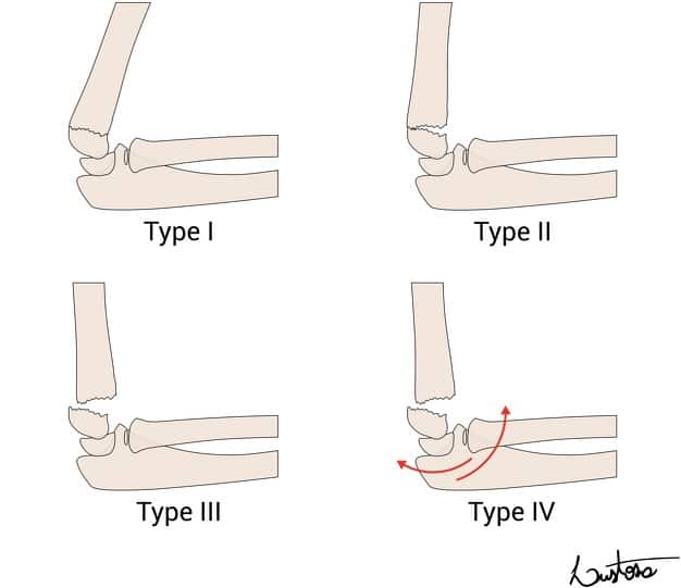
Later subdivisions are used inconsistently. Type I may be subdivided according to medial-column buckling or collapse, and type II is frequently divided into IIA and IIB according to the presence of rotational or translational displacement. Type IIIA and IIIB have been assigned different meanings by different authors and should therefore be defined whenever used.
Historical context
Although bearing Gartland’s name, the modern classification was not created by one author. Earlier displacement-based systems were published by Lubinus, Felsenreich and Holmberg. Gartland introduced a simple three-group clinical framework in 1959. Wilkins later defined the categories according to cortical contact whilst Leitch added the dynamically unstable type IV injury.
The chronology below traces how these successive observations became the modern modified Gartland classification.
History
1924 – Hans Johann Georg Lubinus (1893–1973), then an assistant at the Surgical University Clinic in Kiel, published Über den Entstehungsmechanismus und die Therapie der suprakondylären Humerusfraktur. Drawing upon 125 clinical cases, follow-up examinations and experimental fracture production he challenged a purely extension–flexion interpretation and emphasised shearing, axial angulation, translation and rotation of the distal fragment.
Historical note: Lubinus later joined the NSDAP (National Socialist German Workers’ Party) and SS, and his Kiel clinic participated in compulsory sterilisation under the Nazi regime. Whether this history contributed to the subsequent neglect of his earlier fracture work is uncertain.
1931 – Felsenreich’s application of the Lubinus classification. Fritz Felsenreich published Kindliche suprakondyläre Frakturen und posttraumatische Deformitäten des Ellenbogengelenkes. He divided fractures into three groups according to displacement based on the work of Lubinus:
- group I — incomplete fracture with no or slight axial angulation;
- group II — definite axial angulation in any direction;
- group III — complete displacement, usually by approximately one bone-width.
Felsenreich applied the classification to a clinical series and emphasised rotation, periosteal injury, vascular compromise and the possibility that partially displaced group II fractures could be unexpectedly unstable.
1945 – Holmberg classification. Lennart Holmberg published a monograph on fractures of the distal humerus in children and divided supracondylar fractures into four grades according to displacement:
- no displacement
- displacement with retained posterior cortical continuity
- displacement with rotation
- complete displacement without fragment contact.
Holmberg’s four-stage progression has similarities to the later modified Gartland system, particularly its separation of rotational displacement from complete loss of cortical contact.
1959 – Gartland classification. John Joseph Gartland (1918-2016) published Management of supracondylar fractures of the humerus in children and grouped extension-type injuries into three broad categories:
- Type I — nondisplaced
- Type II — minimal to moderately displaced
- Type III — severely displaced
Gartland used the three groups to organise treatment, emphasising careful neurological and vascular examination, recognition of instability and avoidance of excessive elbow flexion.
1962 – Jean Lagrange (1920–1996) and Pierre Rigault (1931–2024) described a five-stage radiographic classification of extension-type supracondylar humeral fractures in Les fractures de l’extrémité inférieure de l’humérus de l’enfant. Stages I–IV progressed from an incomplete, undisplaced anterior cortical fracture to complete displacement without cortical contact; stage V represented a higher metaphyseal–diaphyseal fracture. The system became widely used in France and other French-speaking countries.
1984 – Kaye Evan Wilkins (1934–2023) published Fractures and Dislocations of the Elbow Region. He replaced Gartland’s subjective distinction between “moderately” and “severely” displaced fractures with the more objective criterion of posterior cortical contact:
- Type I — undisplaced
- Type II — displaced with an intact posterior cortex
- Type III — completely displaced with no cortical contact
Wilkins further divided completely displaced fractures according to the direction of distal-fragment displacement:
- IIIA — posteromedial
- IIIB — posterolateral
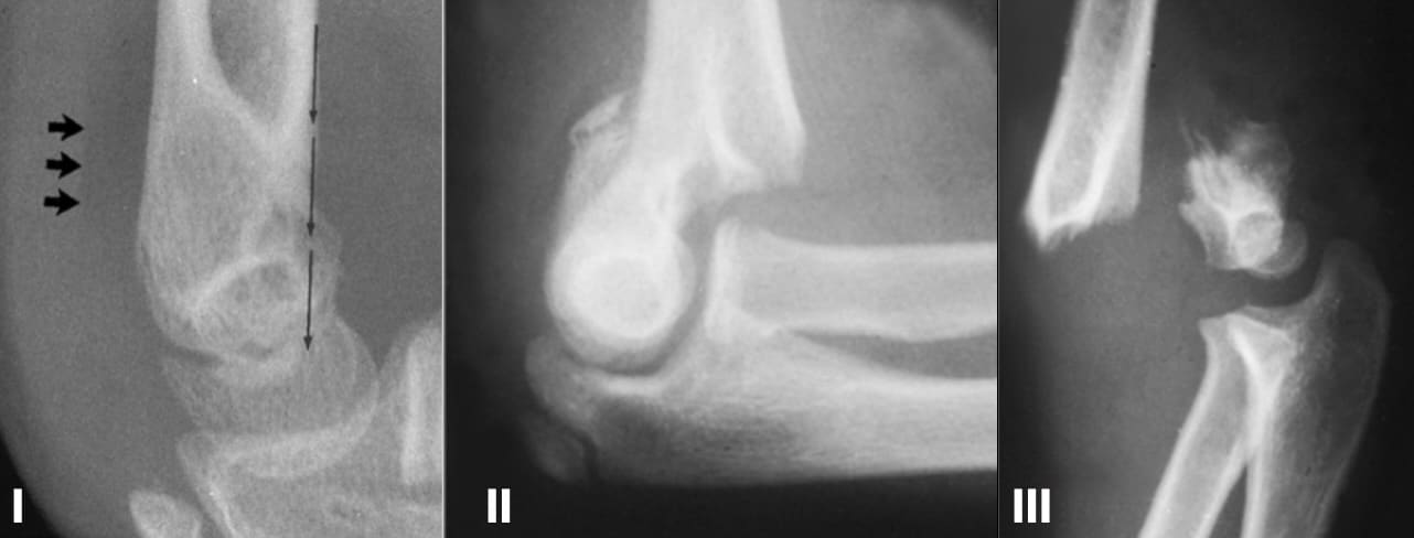
Left: Type I: Undisplaced fracture. The anterior humeral line crosses the ossification centre of the capitellum; displacement of the posterior fat pad supports the presence of an occult fracture.
Middle. Type II: Displaced supracondylar fracture with the posterior cortex intact. Both angulation and rotation of the distal fragment are present.
Right. Type III: Completely displaced fracture with no cortical contact between the fragments.
Wilkins’ published type II example showed both rotation and angulation. The commonly used later subdivision into type IIA fractures without rotation or translation and type IIB fractures with rotational or translational displacement was therefore not part of Wilkins’ classification in the reviewed textbook editions.
1995 – Henri De Boeck and colleagues reported 13 children with supracondylar fractures involving impaction or comminution of the medial column. The injury could appear minimally displaced on the lateral radiograph while loss of the Baumann angle on the anteroposterior view revealed medial-column collapse and a risk of cubitus varus.
This pattern was later incorporated inconsistently into the modified Gartland system as type IB or recorded separately as medial-column comminution.
2001 – Barton and colleagues assessed the reliability of the three-type Wilkins-modified Gartland classification in 50 children. Intraobserver agreement was almost perfect at two weeks and 36 weeks (κ = 0.84 and 0.81), while overall interobserver agreement was substantial (κ = 0.74). Most disagreement occurred between types I and II, the boundary most likely to influence management.
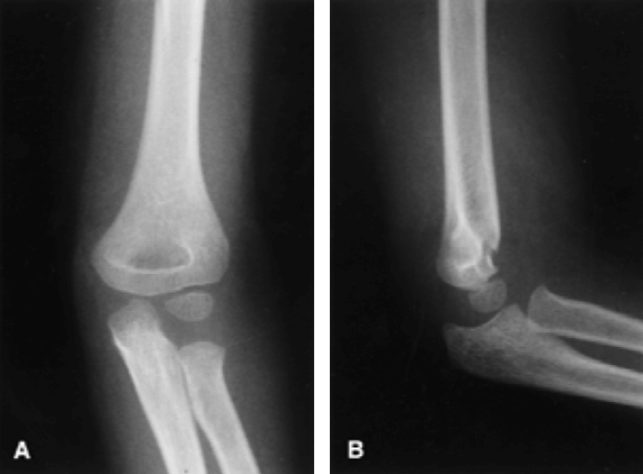
a fracture that caused confusion in classification between type I
and type II supracondylar humerus fractures. Barton 2001
2006 – Leitch and colleagues identified nine multidirectionally unstable fractures among 297 operatively treated supracondylar fractures. Under anaesthesia and fluoroscopy, the distal fragment displaced into both flexion and extension because no functioning periosteal hinge remained. They proposed the designation type IV, noting that instability could result from the original injury or arise during attempted reduction. All nine fractures were treated successfully by closed reduction and percutaneous pinning.
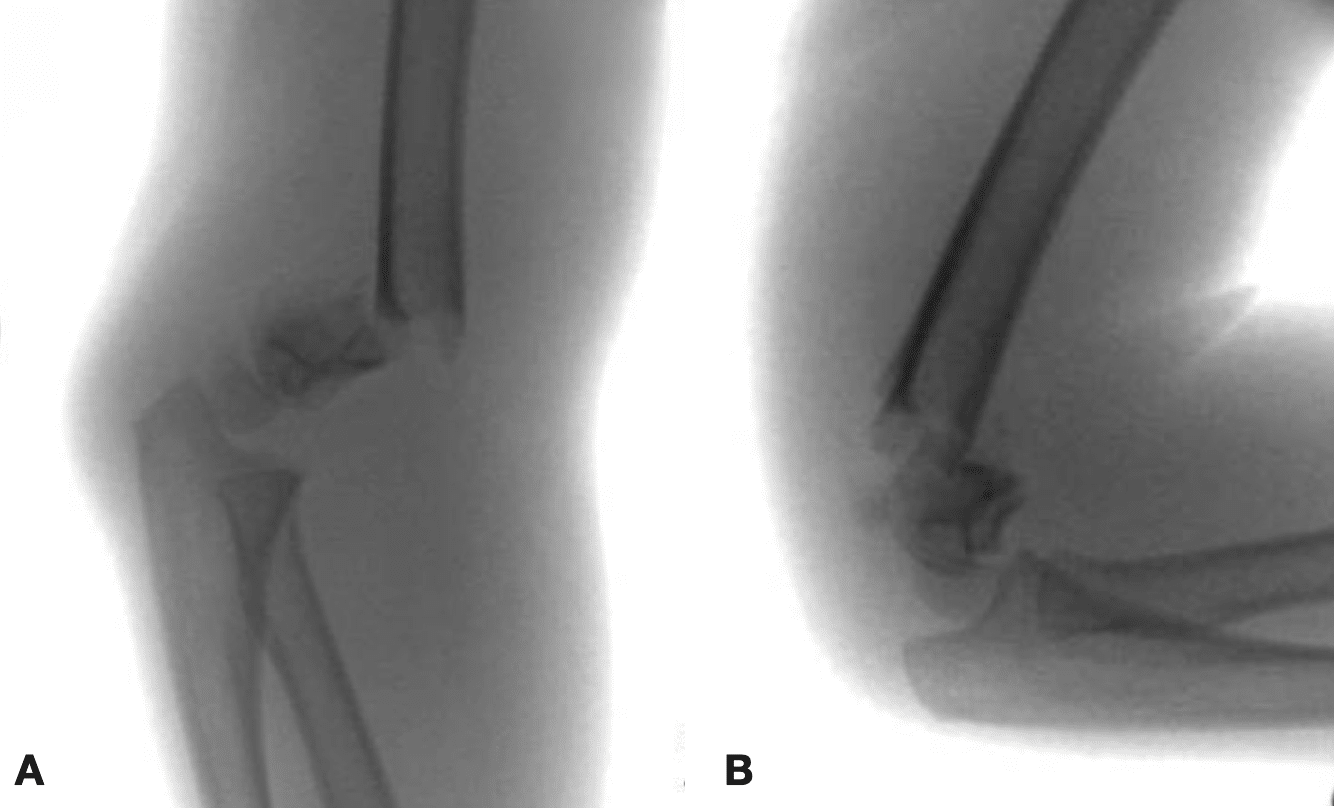
The modern core classification therefore combines Gartland’s three-stage framework, Wilkins’ cortical-contact criteria and Leitch’s dynamically unstable type IV fracture. Later IA/IB, IIA/IIB and IIIA/IIIB subdivisions remain variably defined.
Associated Persons
- Hans Johann Georg Lubinus (1893–1973)
- Fritz Felsenreich (dates unknown)
- Lennart Holmberg (dates unknown)
- John Joseph Gartland (1918-2016)
- Jean Lagrange (1920–1996)
- Pierre Rigault (1931–2024)
- Kaye Evan Wilkins (1934–2023)
References
Historical references
- Lubinus HH. Über den Entstehungsmechanismus und die Therapie der suprakondylären Humerusfraktur. Dtsch Z Chir. 1924; 186: 289–350.
- Felsenreich F. Kindliche suprakondyläre Frakturen und posttraumatische Deformitäten des Ellenbogengelenkes. Arch Orthop Unfallchir. 1931;29:555–578.
- Holmberg L. Fractures in the distal end of the humerus in children. Acta Chir Scand. 1945;92(suppl 103):1–69.
- Gartland JJ. Management of supracondylar fractures of the humerus in children. Surg Gynecol Obstet. 1959;109:145–154
- Lagrange J, Rigault P. Les fractures de l’extrémité inférieure de l’humérus de l’enfant. Rev Chir Orthop, 1962; 48: 334-414
- Wilkins KE. Fractures and Dislocations of the Elbow Region. In: Rockwood CA, Wilkins KE, King R, eds. Fractures in Children. Vol 3. Philadelphia, PA: Lippincott; 1984: 363–575.
- De Boeck H, De Smet P, Penders W, De Rydt D. Supracondylar elbow fractures with impaction of the medial condyle in children. J Pediatr Orthop. 1995 Jul-Aug;15(4):444-8.
- Leitch KK, Kay RM, Femino JD, Tolo VT, Storer SK, Skaggs DL. Treatment of multidirectionally unstable supracondylar humeral fractures in children. A modified Gartland type-IV fracture. J Bone Joint Surg Am. 2006 May;88(5):980-5.
Eponymous term review
- Barton KL, Kaminsky CK, Green DW, Shean CJ, Kautz SM, Skaggs DL. Reliability of a modified Gartland classification of supracondylar humerus fractures. J Pediatr Orthop. 2001 Jan-Feb;21(1):27-30.
- Alton TB, Werner SE, Gee AO. Classifications in brief: the Gartland classification of supracondylar humerus fractures. Clin Orthop Relat Res. 2015 Feb;473(2):738-41.
- Shah M, Agashe MV. Supracondylar Humerus Fractures: Classification Based Treatment Algorithms. Indian J Orthop. 2020 Oct 22;55(1):68-80
- Bridges CS, Taylor TN, Han A, Gladstein AZ, Smith BG, Kan JH. The Gartland classification for expediting supracondylar humerus fracture triage: a collaborative approach to structured reporting between pediatric radiologists and orthopedists. Clin Imaging. 2024 May;109:110118.
eponymictionary
the names behind the name

MBChB - Liverpool. RMO in Emergency Medicine at Sir Charles Gairdner Hospital, currently soaking up the sun down under. Aiming to pursue a career in anaesthetics.

BA MA (Oxon) MBChB (Edin) FACEM FFSEM. Emergency physician, Sir Charles Gairdner Hospital. Passion for rugby, medical history, medical education, and asynchronous learning #FOAMed evangelist. Editor-in-Chief and Webmaster of Life in the Fast lane, LITFL | On Call 4e| Eponyms | Books | Horology |