![]()
Godfrey Hounsfield
Sir Godfrey Newbold Hounsfield (1919-2004) was an English electrical engineer, famous for the invention of computerised tomography (CT) for which he shared the Nobel prize in 1979
Hounsfield was born in Newark, Nottingham; he was the youngest of five children and grew up on a farm. From an early age Godfrey was interested in the machines within the farm he grew up on, undergoing home made dangerous experiments on the farm (including throwing himself off hay stacks with a homemade glider)
He joined the RAF during the second world war where he was taken on as a radar mechanic instructor and moved to the Royal College of Science in South Kensington and then too the Cranwell Radar School. In his spare time at Cranwell he built a oscilloscope and demonstration equipment as aids for people on electronics and radar courses. His work was noticed by Air Vice-Marshall Kennedy who got him a grant to study at the House Electrical Engineering College in London.
The idea for computed topography first came to him in 1967, initially his idea was not related to medicine but a realisation that you could see into a box by taking readings at all angles around it. After this he began working on a system that would be able to take hundreds of X ray beams and create a two dimensional image of tissues. He further developed this idea to record the images on sensors as opposed to an X ray films to create mutliple slices; by placing these slices together it was possible to create a three dimensional image. Hounsfield described this technique in a patent in 1968 and it was granted four years later.
He began practising his technique on cow heads from a slaughterhouse. The first patient to undergo computed tomography was scanned in 1971 in Atkinson Morley hospital with Radiologist James Ambrose. The patient had a suspected cerebral cyst; the scan showed its exact whereabouts and cemented the clinical use of computed tomography.
He received multiple accolades for his work including a Nobel prize in 1979 and in 1981 was knighted. On receiving his Nobel prize he offered the following advice, “Don’t worry if you can’t pass exams, so long as you feel you have understood the subject.” The Hounsfield scale was named after him.
Biography
- Born on August 28, 1919 in Nottinghamshire, England
- DFH (Diploma of Faraday House) from Faraday House Electrical Engineering College in London
- 1949 – Commenced work at Electrical and Musical Instrument (EMI) Ltd
- 1972 – Received the MacRobert award from the Council of Engineering Institutions
- 1975 – Elected a Fellow of the Royal Society (FRS)
- 1976 – Commander of the Order of the British Empire
- 1979 – Nobel Prize in Physiology or Medicine “for the development of computer assisted tomography” with Allan MacLeod Cormack (1924-1998); the first Nobel prize for an invention in diagnostic radiology since Wilhelm Röntgen in 1901
- 1981 – Received a knighthood
- Died on August 12, 2004 in Kingston upon Thames, England
Medical Eponyms
Hounsfield unit (HU)
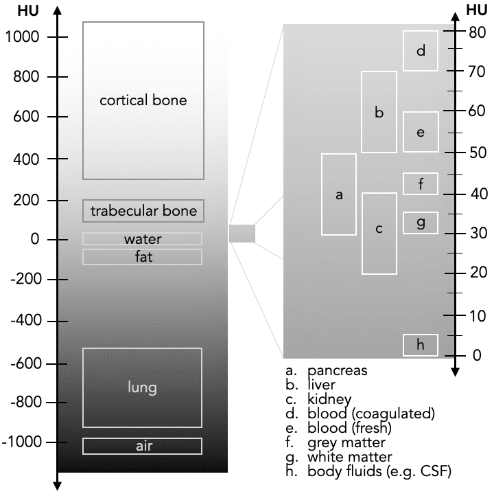
Hounsfield unit (HU) is a relative quantitative measurement of radio density used in the interpretation of computed tomography (CT) images. In the linear scale, the radiodensity of distilled water at a standard temperature and pressure is defined as zero hounsfield units (HU) whereas the radiodensity of air is -1000 (HU). Machines are calibrated using a ‘phantom’ with a known radiodensity, testing whether the machine gives the appropriate HU.
The scale runs from -1000 for air to +2000 for dense bones to over 3000 for metals. Therefore most CT machines and the software for their interpretation of images store values between -1024 to +3071.
Different body tissues and materials have different HU values, such as fat has a value between -120 to -90 and CSF is +15. This helps evaluate different underlying pathologies, such as fat content in the liver and the diagnosis of fatty liver disease. The values can also be used to determine the time scale of certain pathologies; for example subdural haematomas after the first few hours has a value of +75 to +100 but after 10-14 days has a value of +35 to +40.
Major Publications
- Ambrose J, Hounsfield G. Computerized transverse axial tomography. Br J Radiol. 1973 Feb;46(542):148-9
- Hounsfield GN. Computerized transverse axial scanning (tomography). 1. Description of system. Br J Radiol. 1973 Dec;46(552):1016-22
- Hounsfield GN. Picture quality of computed tomography. AJR Am J Roentgenol. 1976 Jul;127(1):3-9
- Hounsfield GN. Historical notes on computerized axial tomography. J Can Assoc Radiol. 1976 Sep;27(3):135-42
- Sagel SS, Weiss ES, Gillard RG, Hounsfield GN, Jost GT, Stanley RJ, Ter-Pogossian MM. Gated computed tomography of the human heart. Invest Radiol. 1977 Nov-Dec;12(6):563-6.
- Hounsfield GN. The E.M.I. scanner. Proc R Soc Lond B Biol Sci. 1977 Jan 14;195(1119):281-9
- Hounsfield GN. Potential uses of more accurate CT absorption values by filtering. AJR Am J Roentgenol. 1978 Jul;131(1):103-6.
- Hounsfield GN. Computed medical imaging. Nobel lecture, December 8, 1979. J Comput Assist Tomogr. 1980 Oct;4(5):665-74
- Hounsfield GN. Computed medical imaging. Science. 1980 Oct 3;210(4465):22-8
References
Biography
- Godfrey N. Hounsfield – Biographical. NobelPrize.org 1979
- Richmond C. Sir Godfrey Hounsfield. BMJ. 2004 Sep 18; 329(7467): 687
- Oransky I. Sir Godfrey N. Hounsfield. Lancet. 2004 Sep 18-24;364(9439):1032.
- Wright P. Obituary: Sir Godfrey Hounsfield. The Guardian. 2004.
- Sherwood I. In Memoriam: Sir Godfrey Hounsfield. Radiology 2005; 234(3): 975-976. [European Radiology (2004; 14: 2152–2153)]
- Wells PNT. Sir Godfrey Newbold Hounsfield KT CBE. Biogr. Mems Fell. R. Soc. 2005; 51: 221-235
- Bates S, Beckmann L, Thomas A, Waltham R. Godfrey Hounsfield: Intuitive Genius of CT. London, UK: The British Institute of Radiology; 2012
- Bhattacharyya KB. Godfrey Newbold Hounsfield (1919-2004): The man who revolutionized neuroimaging. Ann Indian Acad Neurol. 2016 Oct-Dec;19(4):448-450
- Nicholls M. Sir Godfrey Newbold Hounsfield and Allan M. Cormack. Eur Heart J. 2019 Jul 1;40(26):2101-2103
- Thomas AMK, Busch U. Sir Godfrey Hounsfield at 100. Z Med Phys. 2019 Dec;29(4):299-301.
- Bibliography. Hounsfield, Godfrey. WorldCat Identities
Eponymous terms
- Beckmann EC. CT scanning the early days. Br J Radiol. 2006 Jan;79(937):5-8.
- Petrik V, Apok V, Britton JA, Bell BA, Papadopoulos MC. Godfrey Hounsfield and the dawn of computed tomography. Neurosurgery. 2006 Apr;58(4):780-7
- Glide-Hurst C, Chen D, Zhong H, Chetty IJ. Changes realized from extended bit-depth and metal artifact reduction in CT. Med Phys. 2013 Jun;40(6):061711
- de Alcântara ACS, Assis I, Prada D, Mehle K, Schwan S, Costa-Paiva L, Skaf MS, Wrobel LC, Sollero P. Patient-Specific Bone Multiscale Modelling, Fracture Simulation and Risk Analysis-A Survey. Materials (Basel). 2019 Dec 24;13(1):106
- DenOtter TD, Schubert J. Hounsfield Unit. In: StatPearls [Internet]. 2021
Eponym
the person behind the name

Doctor currently working in South Wales, training in anaesthetics. Graduated Leeds University with MB ChB with BSc in microbiology in relation to medicine. Special interests in emergency medicine, critical care and anaesthetics

Assistant Professor of Abdominal Imaging and Intervention at the University of Wisconsin Madison School of Medicine and Public Health. Interests include resident and medical student education, incorporating the latest technology for teaching radiology. I am also active as a volunteer teleradiologist for hospitals in Peru and Kenya. | Medmastery | Radiopaedia | Website | Twitter | LinkedIn | Scopus