Gustav Asboe-Hansen

Gustav Asboe-Hansen (1917-1989) was a Danish dermatologist
Asboe-Hansen conducted internationally recognized research on the biochemistry, structure and function of connective tissue, as well as changes therein in connective tissue diseases, especially scleroderma.
He is eponymously remembered for his description of ‘blister spread’ with application of vertical pressure to a bulla as a clinical sign pertaining to the pemphigus group of diseases (Asboe-Hansen sign).
Biography
- Born on August 12, 1917
- 1951 – MD
- 1949-1960 Head of the connective tissue laboratory at the University of Copenhagen, Department of Anatomy
- 1960-1987 Professor of dermatovenerology (skin and venereal diseases) at the University of Copenhagen and consultant at Rigshospitalet’s department of dermatovenerology
- Died on December 2, 1989
Medical Eponyms
Asboe-Hansen sign
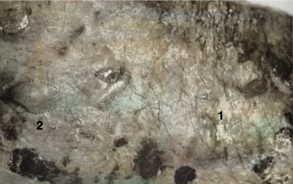
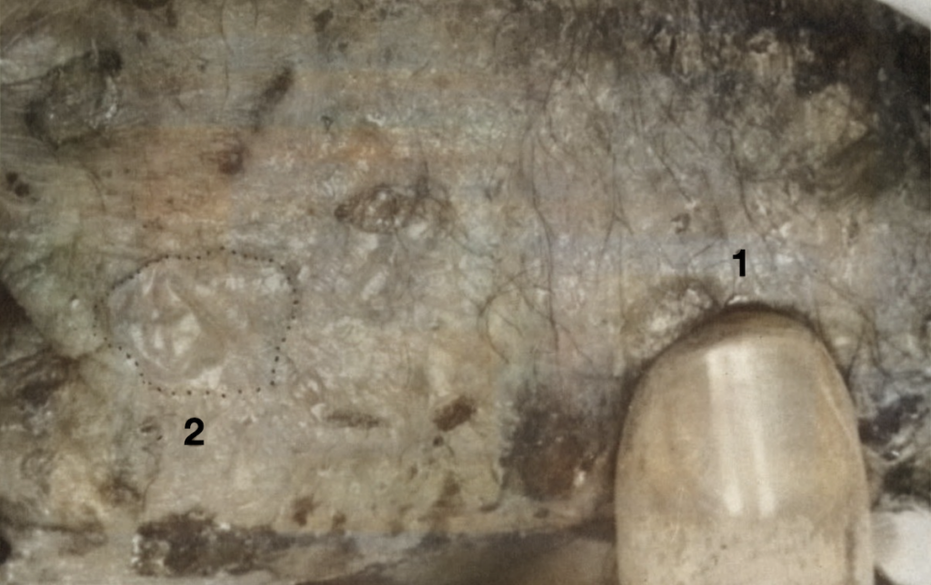
The Asboe-Hansen sign refers to the lateral extension of a bulla to adjacent unblistered skin when vertical pressure is applied.
It is often synonymously used interchangeably to describe the same phenomenon with the terms “Lutz sign,” “Bulla-Spread sign,” “indirect Nikolsky sign” or “Nikolsky II sign”. Lutz was indeed the first to describe the phenomenon in 1957, however referred to its utility specifically in the context of bullous pemphigoid (pemphigus vulgaris chronicus). Asboe-Hansen expanded on this in 1960 and was the first to describe the utility of this test in a number of the pemphigus group of diseases, and noted that it should be employed to generate a fresh bulla for lesional skin biopsy in the evaluation of such disorders.
A spreading of blisters can be artificially induced by slight to moderate external finger pressure in pemphigus vulgaris (acutus), pemphigus foliaceus, pemphigus vegetans and bullous pemphigoid (pemphigus vulgaris chronicus). The sign is characteristic, although possibly not absolutely specific, of the pemphigus group of diseases.
It reflects a reduction or loss of intercellular bridges between epidermal cells or a dermalepidermal lysis. In the new area of the expanded blister, the microscopic picture of a spontaneous fresh blister is reproduced.
Asboe-Hansen 1960


Today, the Asboe-Hansen sign is sometimes considered a subclass of the Bulla-Spread sign, applying specifically to smaller, intact, tense bullae. It is unclear where this distinction originated.
Asboe-Hansen disease
Asboe-Hansen Disease was originally described as a congenital disturbance occurring predominantly in female newborns, characterised by irregularly shaped pigmented macules as well as defects of the integumentary system.
This was described by Asboe-Hansen in 1953 in a report of 4 cases:

On the fourth day of life, red patches of exanthema appeared on the legs and left arm, developing within a few days into vesicles and bullae. “
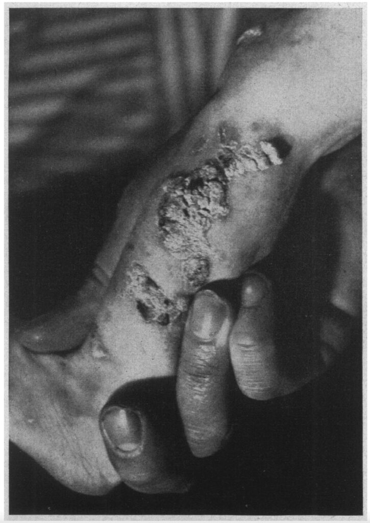
As early as 13 days after the first eruption, it was noted that the skin over the ankles was of a leathery thickness. Thirty-two days later, thick verrucous keratotic outgrowths were noted at the sites of the healed vesicles, particularly on both legs, in the region of the knee, and on the feet.
Asboe-Hansen, 1953
It is now known that Asboe-Hansen was likely describing the early stages of what is today known as “Incontinentia pigmenti,” an X-linked dominant disorder. Incontinentia pigmenti is usually lethal before birth in males. In affected females, it causes highly variable abnormalities of the skin, hair, nails, teeth, eyes, and central nervous system. Cutaneously, it traditionally exhibits stages of vesicular, then verrucous, hyperpigmented and finally atrophic/hypopigmented appearance.
Major Publications
- Asboe-Hansen G. Bullous keratogenous and pigmentary dermatitis with blood eosiniophilia in newborn girls: report of four cases. Archives of Dermatology and Syphilology, Chicago, 1953; 67: 152-157. [Asboe-Hansen disease] [Bloch-Sulzberger pigment dermatosis]
- Asboe-Hansen G. Blister-spread induced by finger-pressure, a diagnostic sign in pemphigus. J Invest Dermatol. 1960 Jan ;34: 5-9. [Asboe-Hansen sign]
- Asboe-Hansen G. Diagnosis of pemphigus. Br J Dermatol. 1970;83:Suppl:81-92.
- Asboe-Hansen G. Blister spreading, a diagnostic test in pemphigus. Loss of epidermal and dermoepidermal cohesion. Arch Dermatol Res. 1988;280 Suppl:S66-7.
References
Biography
- Bibliography. Asboe-Hansen, Gustav. WorldCat Identities
Eponymous terms
- Grando SA, Grando AA, Glukhenky BT, Doguzov V, Nguyen VT, Holubar K. History and clinical significance of mechanical symptoms in blistering dermatoses: a reappraisal. J Am Acad Dermatol. 2003 Jan;48(1):86-92.
- Ganapati S. Eponymous dermatological signs in bullous dermatoses. Indian J Dermatol. 2014 Jan;59(1):21-3
- Dowling JR, Anderson KL, Huang WW. Asboe-Hansen sign in toxic epidermal necrolysis. Cutis. 2019 Apr;103(4):E6-E8.
- Bartolucci SL. Stedman’s Medical Eponyms.2nd ed. Philadelphia: Lippincott, Williams & Wilkins; 2005. p. 29.
- Ganapati S. Eponymous Dermatological Signs in Bullous Dermatoses. Indian Journal of Dermatology. 2014 59(1):21-23.
[cite]
BSc, MD from University of Western Australia. Junior Doctor currently working at Sir Charles Gairdner Hospital.

