
![]()
Hydropneumothorax
A young woman presented with 2 days shortness of breath and right sided chest discomfort after a long haul flight. What does this scan demonstrate?
Reveal Answer
- This image is taken at the costophrenic angle.
- The curved diaphragm is seen moving up and down with shallow respiration. There is a small associated pleural effusion. Above this is pneumothorax.
- The moving air / fluid level may be mistaken for lung sliding or a lung point, however look at the pleural surface cranial to the junction between fluid and air. There is no sliding, just the static typical appearance of pneumothorax.
Ultrasound of hydropneumothorax explained
A second scan is performed on the same patient with the probe on the anterior chest wall. What does this scan demonstrate?
Reveal Answer
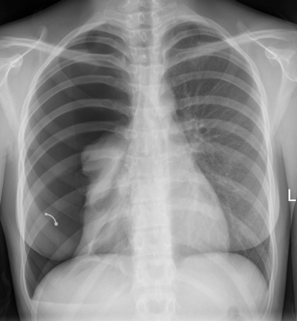
- There is a large right sided pneumothorax (confirmed on CXR as below)
- The horizontal air fluid level seen at the level of the right diaphragm is the small associated pleural effusion.
- A nipple piercing is also present.
Related Clinical Cases
- LITFL Ultrasound library
- LITFL Top 100 ultrasound cases
- LUNG ultrasound cases
- LUNG ultrasound modules

ULTRASOUND LIBRARY
Modules

An Emergency physician based in Perth, Western Australia. Professionally my passion lies in integrating advanced diagnostic and procedural ultrasound into clinical assessment and management of the undifferentiated patient. Sharing hard fought knowledge with innovative educational techniques to ensure knowledge translation and dissemination is my goal. Family, wild coastlines, native forests, and tinkering in the shed fills the rest of my contented time. | SonoCPD | Ultrasound library | Top 100 | @thesonocave |

¿No debería el punto pulmón corresponder a una zona lateral y no tan alto debido a su tamaño?