


Iselin disease
Iselin disease is a painful traction apophysitis of the fifth metatarsal tuberosity in skeletally immature children and adolescents. The apophysis lies at the insertion of the peroneus brevis tendon and becomes symptomatic through repetitive traction and overuse, particularly with running, jumping and inversion-related activity.
Typical presentation is activity-related lateral foot pain, often with focal tenderness and prominence or swelling over the base of the fifth metatarsal. Pain may be aggravated by running, jumping, footwear pressure and resisted eversion. A preceding traumatic event is often absent, although symptoms may begin after a minor inversion injury
Investigations
Oblique radiographs are the most useful plain-film projection because the apophysis is inferolateral and may be poorly demonstrated on AP or lateral views. The normal apophysis is oriented longitudinally, parallel to the long axis of the fifth metatarsal. In Iselin disease it may become enlarged, fragmented, rounded or irregular, with widening or ondulation of the apophyseal line.
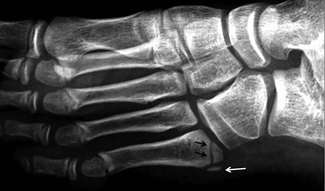
MRI is not routinely required, but when performed may show bone marrow oedema within the apophysis and adjacent metatarsal base with surrounding soft-tissue oedema.
Differential diagnosis
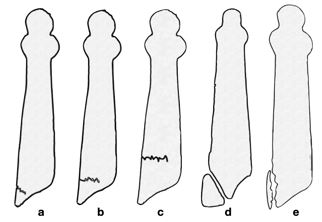
The principal differential diagnoses are avulsion fracture, Jones fracture, proximal fifth-metatarsal stress fracture, os vesalianum pedis and a normal developmental apophysis. The key distinction on X-ray is orientation. The apophyseal line is usually longitudinal, whereas avulsion and Jones-type fractures are usually transverse or oblique.
Treatment is usually conservative, with activity modification, rest, analgesia, ice and footwear modification or padding. Short-leg immobilisation can be used for more symptomatic cases. Prognosis is generally good, with symptoms resolving as the apophysis matures and fuses. In the largest published clinical series, all 27 patients healed, with a mean return to sport of 38 days.
History
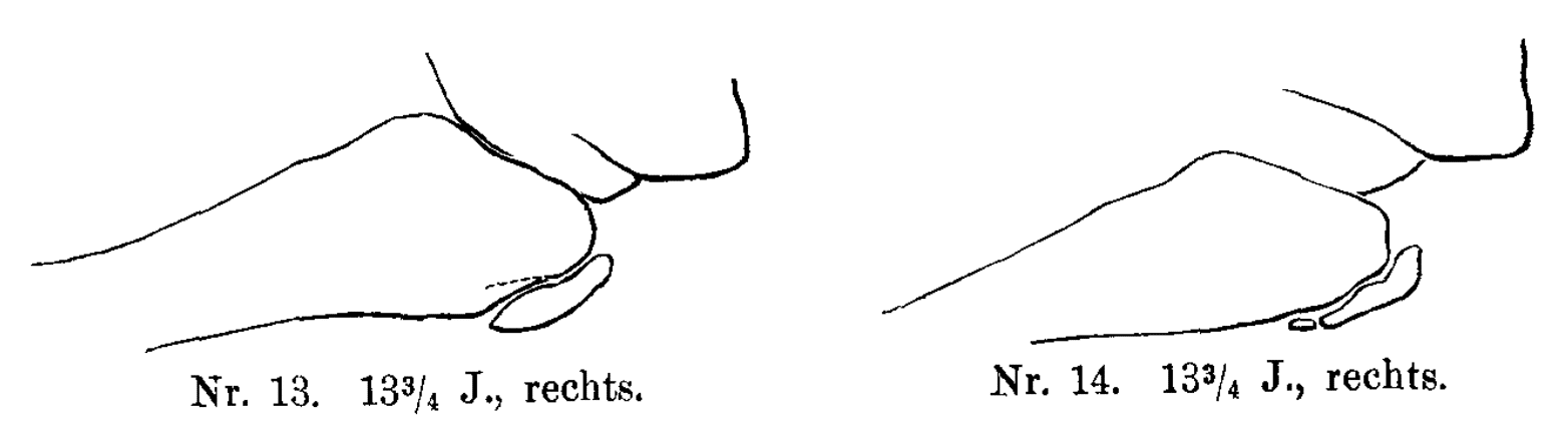
1885 – Wenzel Leopold Gruber (1814-1890) investigated the normal secondary ossification centre of the fifth-metatarsal tuberosity. After observing a separate ossification centre on radiographs of two 13-year-olds, he examined approximately 25 healthy boys aged 12–16 years and concluded that it was probably a normal, near-constant developmental structure, most apparent around age 13¾ years.
1903 – Carl Bernhard Schlatter (1864-1934) described the painful adolescent disorder of the tibial tuberosity initially interpreted as an incomplete avulsion fracture. By 1908 he questioned whether these represented avulsion fractures or growth abnormalities.
1908 – Hans Iselin (1878-1953) published Die Wenzel Grubersehe fibulare Epiphyse der Tuberositas metatarsi quinti. Iselin investigated the normal secondary ossification centre at the fifth-metatarsal tuberosity. He studied 25 healthy boys aged 12–16 years and concluded that it was probably a normal, near-constant developmental structure, most apparent around age 13¾ years.
Iselin warned that the normal apophysis could be mistaken for a fracture. He gave Gruber priority and called it the Wenzel-Gruber fibular epiphysis
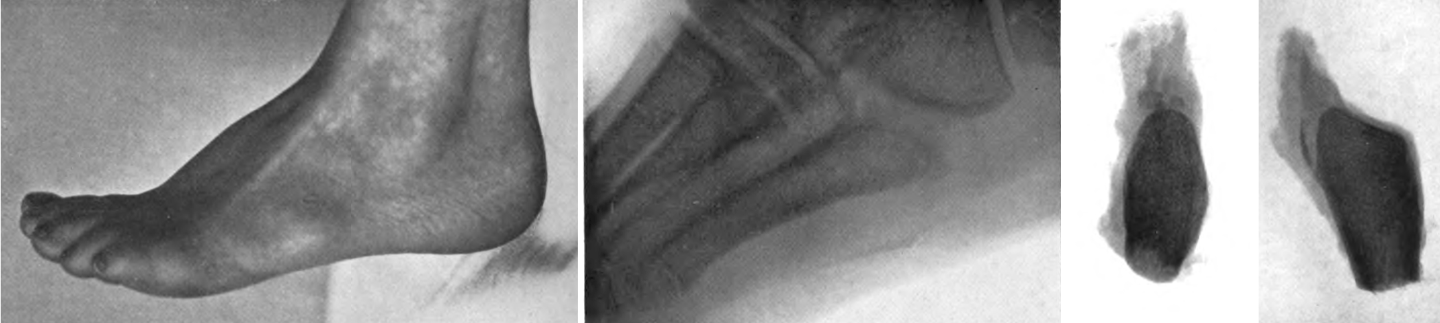
1912 – Iselin published published Wachstumsbeschwerden zur Zeit der knöchernen Entwicklung der Tubersositas metatarsi quinti. His described a disease of the fifth-metatarsal tuberosity corresponding closely to Schlatter disease of the tibial tuberosity, adding that the condition had not previously been described.
Case 1 (1909): 13-year-old girl attended the surgical polyclinic with painful swelling over the fifth-metatarsal tuberosity. The area was firm and tender and painful during walking, but there was no erythema, oedema or history of trauma. Radiographs showed the ossification centre to be larger on the symptomatic side.
Iselin did not describe the pathophysiology of traction apophysitis but concluded:
Development of apophyses at tendon insertions may be accompanied by pain and swelling, at the fifth-metatarsal tuberosity just as at the tibial tuberosity, without preceding injury…a sufficient explanation for this painful development of the apophysis cannot yet be given
1986 – Richard C. Lehman, John R. Gregg and Elisabeth Torg reported Iselin’s disease in young athletes and emphasised repetitive athletic stress and inversion as precipitating factors. They proposed recurrent microtrauma and repetitive traction of the peroneus brevis at the insertional apophysis as the underlying mechanism, particularly during running, jumping and inversion stress.
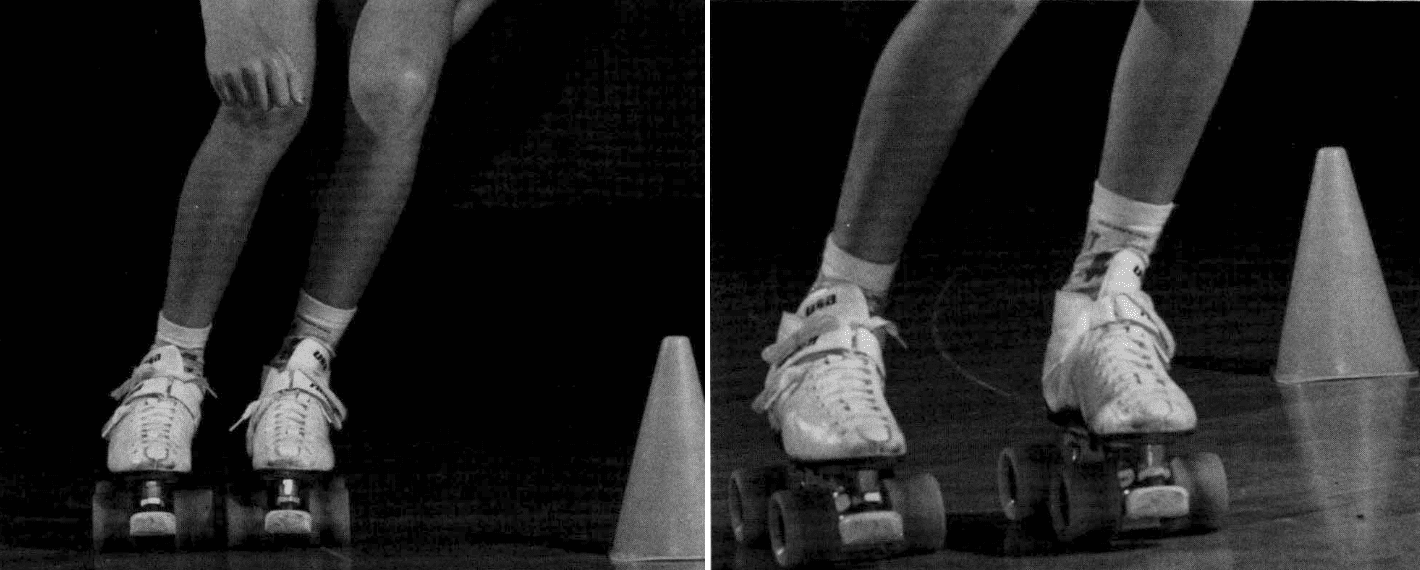
1992 – S. Terry Canale and Keith D. Williams reported four patients with Iselin’s disease and provided a more detailed clinicoradiographic description. They emphasised tenderness over the prominent fifth-metatarsal base, pain with resisted eversion, and the value of the oblique radiograph. They also clarified that the apophyseal line runs parallel to the fifth-metatarsal shaft, helping distinguish it from transverse avulsion fractures.
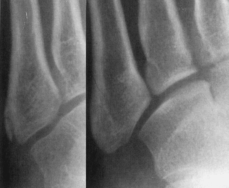
2014 – Deniz et al published Traction apophysitis of the fifth metatarsal base in a child: Iselin’s disease. They presented a symptomatic 10-year-old girl and summarised the concept of Iselin disease as a traction apophysitis. They highlighted oblique radiography, fragmentation and irregularity of the apophysis, and the importance of distinguishing it from fractures and os vesalianum.
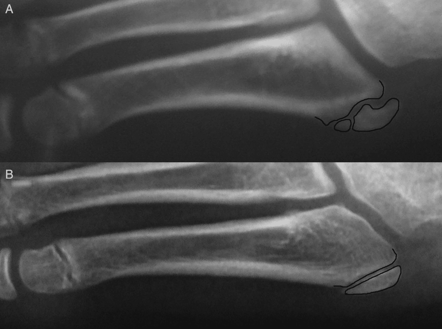
2015 – Sylvester and Hennrikus reported 27 adolescent athletes, the largest clinical series to date. Six had initially been misdiagnosed, including five as fractures. The same diagnostic pitfall Iselin had warned about in 1908. All healed with conservative treatment; with a mean return to sport was 38 days.
2016 – Kishan et al demonstrated marrow and surrounding soft-tissue oedema on MRI, while emphasising that MRI is usually unnecessary when clinical and radiographic findings are typical.
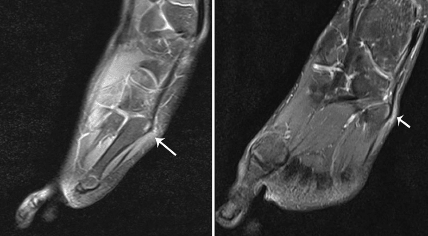
2017 – Forrester et al. reviewed the literature and concluded that Iselin disease is probably under-recognised, emphasising repetitive traction and differentiation from fracture and symptomatic os vesalianum.
Eponymythology
Iselin deserves the eponymous term for the symptomatic clinical disorder, but not for discovery of the fifth-metatarsal apophysis. Wenzel Gruber described the anatomical epiphysis in 1885 and Iselin acknowledged his priority. Iselin’s contribution was to recognise that this normally developing apophysis could become a distinct painful clinical syndrome and to describe it in 1912.
“Iselin disease” is later terminology. Iselin did not name a disease after himself and did not even call it apophysitis. His title was essentially “growth complaints during the bony development of the fifth-metatarsal tuberosity.” The modern pathological term traction apophysitis is a retrospective interpretation.
Associated Persons
- Wenzel Leopold Gruber (1814-1890)
- Hans Iselin (1878-1953)
- Carl Bernhard Schlatter (1864-1934)
Alternative names
- Iselin disease, Iselin’s disease
- Fifth-metatarsal apophysitis, traction apophysitis of the fifth metatarsal, apophysitis of the fifth-metatarsal base.
- Traction apophysitis
References
Historical references
- Gruber W. Auftreten der Tuberositas des Os metatarsale 5, sowohl als persistierende Epiphyse, als auch mit einer an ihrem äußeren Umfange aufsitzenden persistierenden Epiphyse. Archiv für pathologische Anatomie und Physiologie und für klinische Medizin, 1885; 99: 460-471
- Schlatter C. Verletzungen des schnabelförmigen Forsatzes der oberen Tibiaepiphyse. Beiträge zur klinischen Chirurgie, 1903; 38: 874-87
- Schlatter C. Unvollständige Abrißfrakturen der Tuberositas tibiae oder Wachstumsanomalien? Bruns’ Beiträge zur klinischen Chirurgie 1908; 59: 518
- Iselin H. Die Wenzel Grubersehe fibulare Epiphyse der Tuberositas metatarsi quinti im Röntgenbild. Ein wahrscheinlich konstanter Befund im 13. oder 14. Lebensjahre. Deutsche Zeitschrift für Chirurgie 1908; 92: 561-568
- Iselin H. Wachstumsbeschwerden zur Zeit der knöchernen Entwicklung der Tubersositas metatarsi quinti. Deutsche Zeitschrift für Chirurgie 1912; 117: 529–535.
Eponymous term review
- Lehman RC, Gregg JR, Torg E. Iselin’s disease. Am J Sports Med. 1986 Nov-Dec;14(6):494-6.
- Canale ST, Williams KD. Iselin’s disease. J Pediatr Orthop. 1992 Jan;12(1):90-3.
- Deniz G, Kose O, Guneri B, Duygun F. Traction apophysitis of the fifth metatarsal base in a child: Iselin’s disease. BMJ Case Rep. 2014 May 15;2014:bcr2014204687.
- Sylvester JE, Hennrikus WL. Treatment outcomes of adolescents with Iselin’s apophysitis. J Pediatr Orthop B. 2015 Jul;24(4):362-5.
- Kishan TV, Mekala A, Bonala N, Sri Pavani B. Iselin’s disease: Traction apophysitis of the fifth metatarsal base, a rare cause of lateral foot pain. Med J Armed Forces India. 2016 Jul;72(3):299-301.
- Forrester RA, Eyre-Brook AI, Mannan K. Iselin’s Disease: A Systematic Review. J Foot Ankle Surg. 2017 Sep-Oct;56(5):1065-1069
eponymictionary
the names behind the name

Sujit Iyer, MD, is the National Director of Pediatric Services at US Acute Care Solutions and a Professor of Pediatrics and Pediatric EM Fellowship Director at UT Austin Dell Medical School. He completed his medical training and pediatrics residency at Baylor College of Medicine / Texas Children’s Hospital, followed by a Pediatric Emergency Medicine fellowship at Children’s Hospital of Philadelphia. Dr. Iyer focuses on emergency department operations, pediatric readiness in community hospitals, and curriculum development, lecturing nationally on quality improvement and pediatric emergency medicine best practices.

BA MA (Oxon) MBChB (Edin) FACEM FFSEM. Emergency physician, Sir Charles Gairdner Hospital. Passion for rugby, medical history, medical education, and asynchronous learning #FOAMed evangelist. Editor-in-Chief and Webmaster of Life in the Fast lane, LITFL | On Call 4e| Eponyms | Books | Horology |