


Klein line
Klein line is a radiographic reference used on the AP pelvis or hip radiograph to aid the diagnosis of slipped capital femoral epiphysis (SCFE). It is drawn by extending the straight superior cortical margin of the femoral neck proximally.
In the normal growing hip, the line transects a portion of the lateral capital femoral epiphysis. With SCFE, it may intersect less of the epiphysis than expected or, with greater displacement, fail to intersect it altogether.
On the AP radiograph, Klein line demonstrates the medial component of epiphyseal displacement. A normal-appearing Klein line does not exclude SCFE, particularly when slipping is predominantly posterior. Appropriate lateral imaging remains essential when clinical suspicion persists.
Modern teaching
Klein line is commonly taught as a binary rule. The Klein line should intersect the capital epiphysis, and failure to intersect suggests SCFE. This is useful shorthand, but it represents an over simplification of Klein’s original description. See Eponymythology below.
Original description
Klein’s group first established what the normal growing hip should look like. In their 1952 publication they noted that the femoral head normally overhangs the neck so that proximal extension of the straight superior neck margin transects part of the head.
For early medial slipping they described two abnormal possibilities:
failure of the prolonged superior neck line to transect as much of the head as in the ‘normal’ hip
and, with more obvious displacement:
prolongation of the superior neck line proximally will not transect the head.
Therefore, reduced intersection itself was considered abnormal and complete non-intersection was not required for the diagnosis.
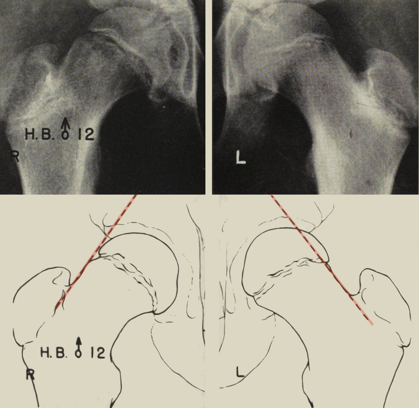
History of the Klein line
Development of the radiographic method
1950–1952 – Armin Klein (1892–1954) and colleagues presented their radiographic studies of SCFE to the American Roentgen Ray Society in September 1950 and the American Academy of Orthopaedic Surgeons in January 1951.
The 1951 AJR publication Roentgenographic features of slipped capital femoral epiphysis emphasised standardised bilateral AP and lateral imaging and comparison with normal growing hips.
In the 1952 JBJS paper Slipped capital femoral epiphysis; early diagnosis and treatment facilitated by normal roentgenograms they described proximal prolongation of the superior femoral-neck line. They noted that early slipping caused it to transect less of the head than normally, or not transect it at all.
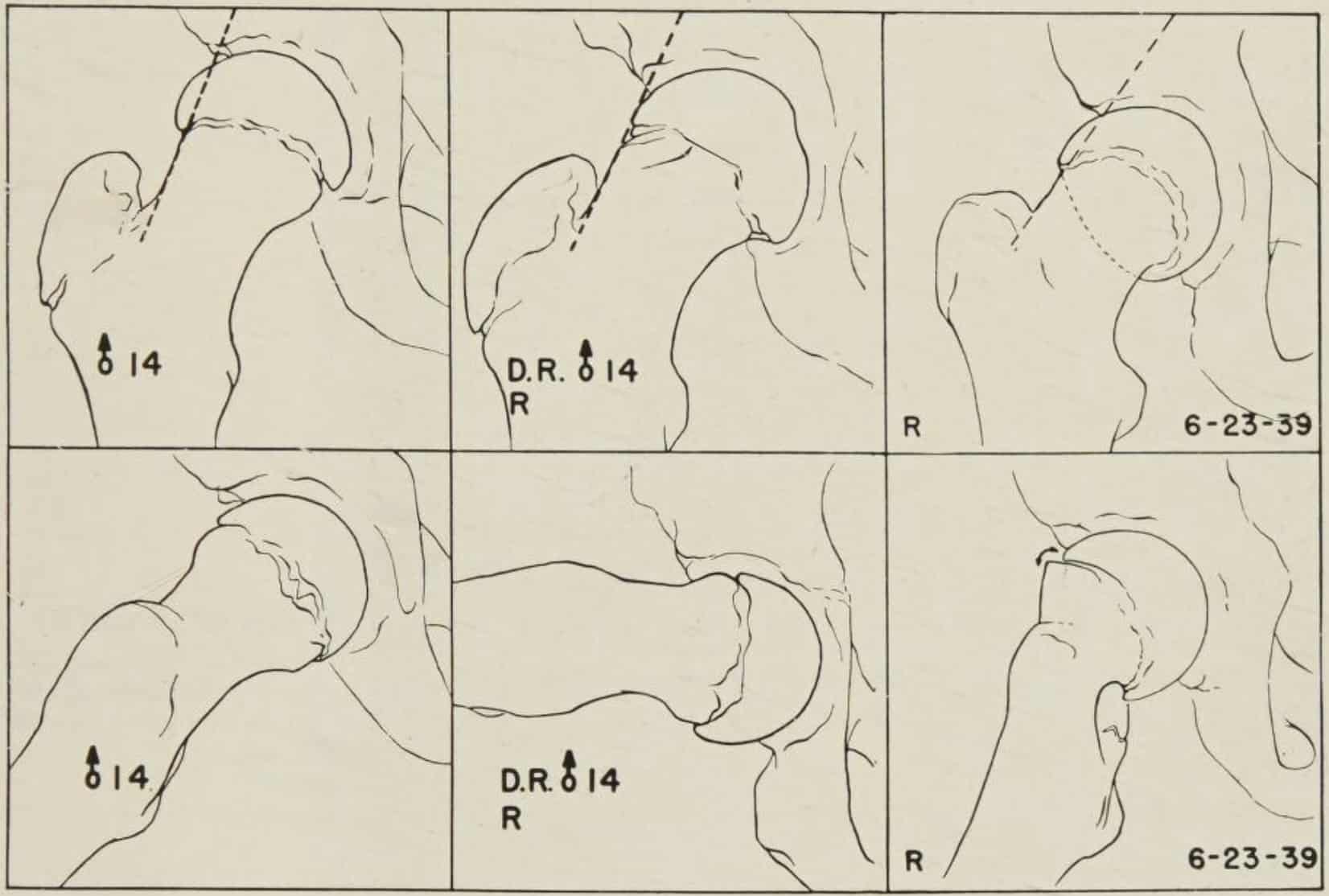
1953 – Klein et al published Slipped Capital Femoral Epiphysis in which they consolidated the technique and clarified the normal anatomy underlying the sign. Klein again emphasised the straight proximal portion of the superior femoral-neck margin and described both reduced and absent intersection with the capital epiphysis.
2009 – Green and colleagues tested the commonly used binary definition of Klein line (complete failure to intersect the epiphysis), and found a sensitivity of 40.3% in predominantly mild unilateral slips. Green introduced a quantitative modification by measuring the width of epiphysis lateral to Klein line and using a ≥2 mm side-to-side difference which increased sensitivity to 79%.
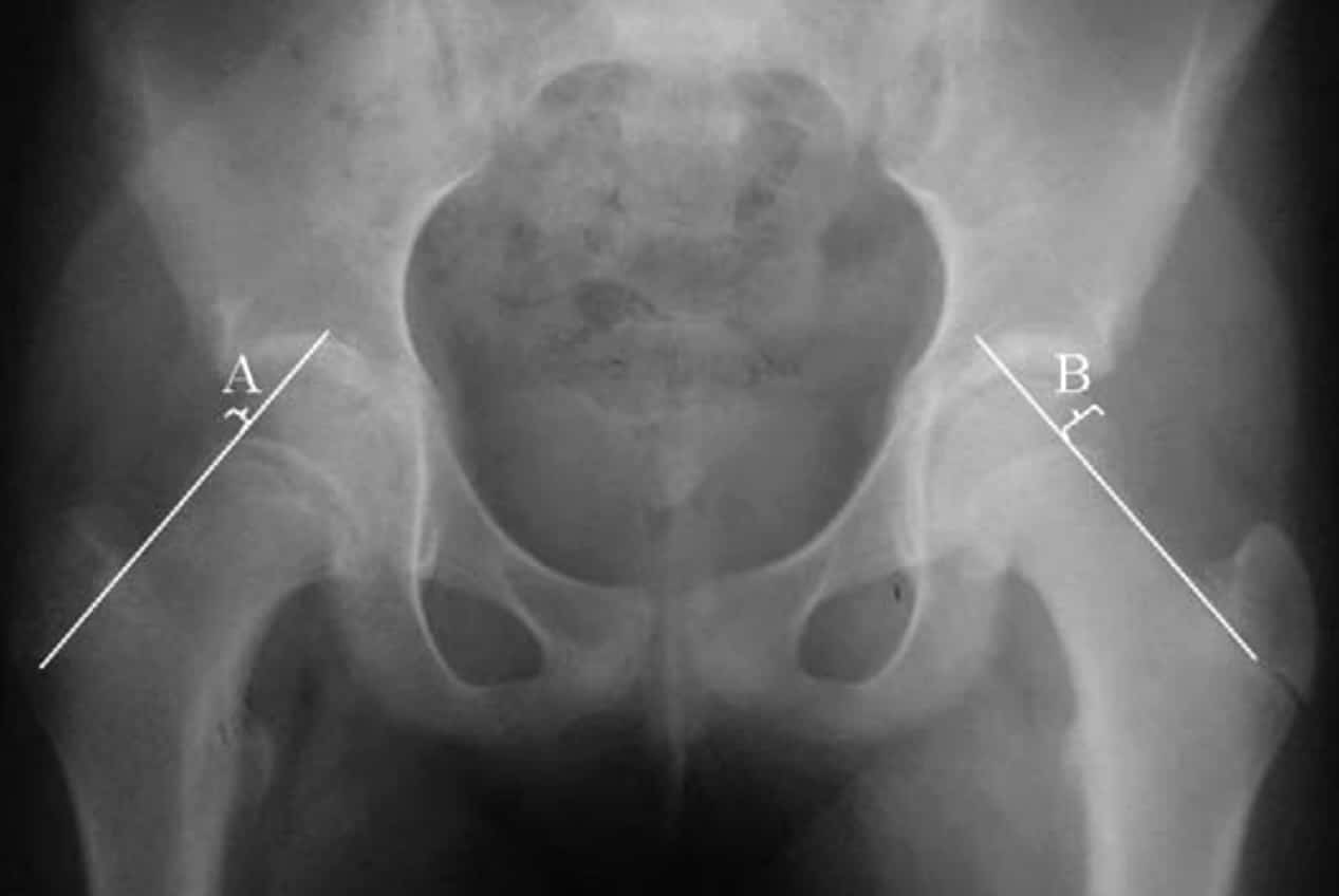
A normal Klein line therefore does not exclude SCFE. Appropriate lateral imaging remains important when the diagnosis is clinically suspected.
Eponymythology
From comparative observation to binary sign
Modern descriptions define a positive Klein sign as complete failure of the line to intersect the capital femoral epiphysis. However, that was only one end of the spectrum described by Klein and colleagues.
Their original method was comparative. In the normal growing hip, proximal extension of the straight superior femoral-neck margin transected part of the head. With early medial slipping, the line might still cross the epiphysis but transect less of it than expected, but with greater displacement it could miss the head entirely.
Klein’s group did not rely on the line in isolation. They advocated standardised bilateral AP and lateral radiographs, comparison with normal hips of similar age and sex, and assessment of additional findings including physeal widening and irregularity, uncovering of the superior femoral neck, altered head-neck contour and posterior displacement on the lateral view.
In 2009, Green and colleagues evaluated what had become the “classic” intersection versus non-intersection Klein line and found only 40.3% sensitivity. Their modified Klein line measured how much epiphysis remained lateral to the line and compared the two hips.
Interestingly, this quantitative modification is similar to Klein’s original description in which the amount of epiphysis transected mattered. Green et al. converted that qualitative observation into a reproducible numerical measurement.
Which comparative hip?
A further limitation concerns the choice of the contralateral hip as the reference standard. Klein’s group discovered substantial occult contralateral involvement and concluded that the opposite hip could not always be assumed to be normal. As the opposite hip could be unreliable as the normal standard, they assembled age- and sex-matched reference radiographs of genuinely normal hips.
Green et al. selected patients whose contralateral hips remained normal on follow-up, allowing side-to-side measurement in their study. In clinical practice, however, bilateral SCFE remains a potential limitation of purely contralateral comparison.
| Klein 1951–1953 | Common modern shorthand | Green et al. 2009 | |
|---|---|---|---|
| Construction | Prolong straight superior femoral-neck margin | Superior femoral-neck line | Same anatomical line |
| Normal | Transects a portion of head/epiphysis | Intersects epiphysis | Measure epiphysis lateral to line |
| Abnormal | Transects less than expected, or none | Non-intersection | ≥2 mm side-to-side difference |
| Reference | Normal developmental anatomy; bilateral films | Often binary AP assessment | Contralateral normal hip |
| Role | One component of broader AP + lateral assessment | Binary AP sign | Quantitative AP measurement |
Related eponym: Trethowan sign
Trethowan sign is a historical British eponym for an abnormal superior femoral-neck relationship in SCFE and is now often used for the abnormal finding produced by Klein line.
No contemporary description by William Henry Trethowan (1882–1934) has been located. The earliest printed attribution currently identified is W. A. Crabbe’s 1968 Guy’s textbook, while Apley later used the term for the familiar superior-neck line failing to pass through the femoral head.
See: Trethowan sign – attribution, historical definitions and relationship to Klein line.
Associated Persons
- Armin Klein (1892–1954)
- William Henry Trethowan (1882–1934)
Alternative names
- Klein’s line
- Line of Klein
Related terms
- Klein sign — positive/abnormal finding
- Trethowan sign — historical British eponym for the abnormal relationship
References
Historical references
- Klein A, Joplin RJ, et al. Roentgenographic changes in nailed slipped capital femoral epiphysis. J Bone Joint Surg Am. 1949 Jan;31A(1):1-22.
- Klein A, Joplin RJ, Reidy JA, Hanelin J. Roentgenographic features of slipped capital femoral epiphysis. Am J Roentgenol Radium Ther. 1951 Sep;66(3):361-74
- Klein A, Joplin RJ, Reidy JA, Hanelin J. Slipped capital femoral epiphysis; early diagnosis and treatment facilitated by normal roentgenograms. J Bone Joint Surg Am. 1952 Jan;34-A(1):233-9.
- Klein A, Joplin RJ, Reidy JA, Hanelin J. Management of the contralateral hip in slipped capital femoral epiphysis. J Bone Joint Surg Am. 1953 Jan;35-A(1):81-7.
- Klein A, Joplin RJ, Reidy JA, Hanelin J. Slipped Capital Femoral Epiphysis. 1953
Eponymous term review
- Burrows HJ. Slipped upper femoral epiphysis; characteristic of a hundred cases. J Bone Joint Surg Br. 1957 Nov;39-B(4):641-58.
- Cowell HR. The significance of early diagnosis and treatment of slipping of the capital femoral epiphysis. Clin Orthop Relat Res. 1966 Sep-Oct;48:89-94.
- Matava MJ, Patton CM, Luhmann S, Gordon JE, Schoenecker PL. Knee pain as the initial symptom of slipped capital femoral epiphysis: an analysis of initial presentation and treatment. J Pediatr Orthop. 1999 Jul-Aug;19(4):455-60
- Green DW, Mogekwu N, Scher DM, Handler S, Chalmers P, Widmann RF. A modification of Klein’s Line to improve sensitivity of the anterior-posterior radiograph in slipped capital femoral epiphysis. J Pediatr Orthop. 2009 Jul-Aug;29(5):449-53.
- Kyle E, Cadogan M. Hip and pelvic lines and arcs. LITFL
eponymictionary
the names behind the name

MB BAO BCh Trinity College Dublin, MSc Clinical Data Analytics University of Galway. Junior doctor in Perth, doctoral researcher in MRI radiomics. Working towards radiology training.

BA MA (Oxon) MBChB (Edin) FACEM FFSEM. Emergency physician, Sir Charles Gairdner Hospital. Passion for rugby, medical history, medical education, and asynchronous learning #FOAMed evangelist. Editor-in-Chief and Webmaster of Life in the Fast lane, LITFL | On Call 4e| Eponyms | Books | Horology |