


Maurice Sokolow

Maurice Sokolow (1911-2002) was an American Cardiologist and educator
Sokolow was known for his contributions to ECG interpretation and hypertension. He is eponymously known for his development of ECG criteria for left ventricular hypertrophy (Sokolow-Lyon criteria), but also made contributions to the development of ambulatory blood pressure measurement.
His work explored the relationship between blood pressure and coronary artery disease; circadian variation in blood pressure as well as contributed to the concept of blood pressure variation and noted that blood pressure recorded during daily life was often lower than that recorded in the Doctor’s office – leading to the concept of “white coat hypertension“
One of his important legacies was his textbook on clinical cardiology which was translated into five languages
Biography
- Born on May 19, 1911 in New York
- 1917 – following the death of his mother at age 6 and with his father unable to care for him, Maurice spent seven years in an orphanage
- graduated UC Berkley
- 1936 – graduated UCSF School of Medicine – recipient of Gold Headed Cane Award (outstanding graduate on his class)
- Residency at the New England Medical Centre (Boston)
- Fellowship in Cardiology at the Michael Reese Hospital (Chicago)
- World War II – served on a hospital ship in the Pacific and developed an interest in tropical diseases
- 1949 – described the ECG characteristics present with left ventricular hypertrophy along with Dr Thomas P Lyon
- 1950s – chief of the hypertension clinic at San Francisco General Hospital Medical Center; founding member of the UCSF Cardiovascular Research Institute
- 1953-1973 chief of cardiology at UC Medical Center
- 1961 – prospective study which developed a portable BP apparatus for automatic blood pressure measurement – initially approaching multiple electronic firms including Hewlett—Packard and Ampex, before working with the Alpha Scientific Corporation and the Remler Company.
- Died from lymphoma on September 26, 2002 at San Francisco, California
Medical Eponyms
Sokolow–Lyon criteria (1949)
Maurice Sokolow and Thomas Lyon tried to improve recognition of early and atypical LVH using the recently introduced unipolar limb and precordial leads. They aimed to define abnormalities seen on the ECG, examine the importance of cardiac position and determine whether delayed ventricular activation provided additional diagnostic information [Sokolow and Lyon, 1949].
Original criterion
SV1 + max(RV5,V6) ≥ 35 mm
The original scheme was broader than this voltage sum and also incorporated lateral precordial voltage, ST–T abnormalities, delayed ventricular activation and cardiac orientation.
Evidence and significance
They selected 200 patients with an abnormal ECG and a clinical condition associated with increased left ventricular load, including hypertension, aortic valve disease and coarctation. After exclusions a group of 147 patients, 90% of whom had severe hypertension, were compared with 151 apparently healthy controls.
The combined SV1+ max(RV5,V6) voltage reached 35 mm in 48 of 147 patients with presumed LVH but in none of the controls. However, this was not an anatomical validation study and LVH was inferred from the clinical condition, ECG and cardiac size rather than measured by echocardiography, cardiac magnetic resonance or autopsy LV mass.
The precordial leads helped identify patients whose standard limb leads appeared normal or “atypical”. The paper reinforced that repolarisation abnormalities and delayed activation could be diagnostically important even when voltage criteria were absent.
Modern interpretation
Today, the Sokolow–Lyon criterion usually refers only to SV1 + max(RV5,V6) ≥ 35 mm
Additional findings from the same paper that remain in use are:
- RV5 or RV6 >26 mm
- RaVL >11 mm
- R-wave peak time >50 ms in V5 or V6
Sokolow–Lyon voltage remains supportive when positive but is too insensitive to exclude anatomical LVH when negative.
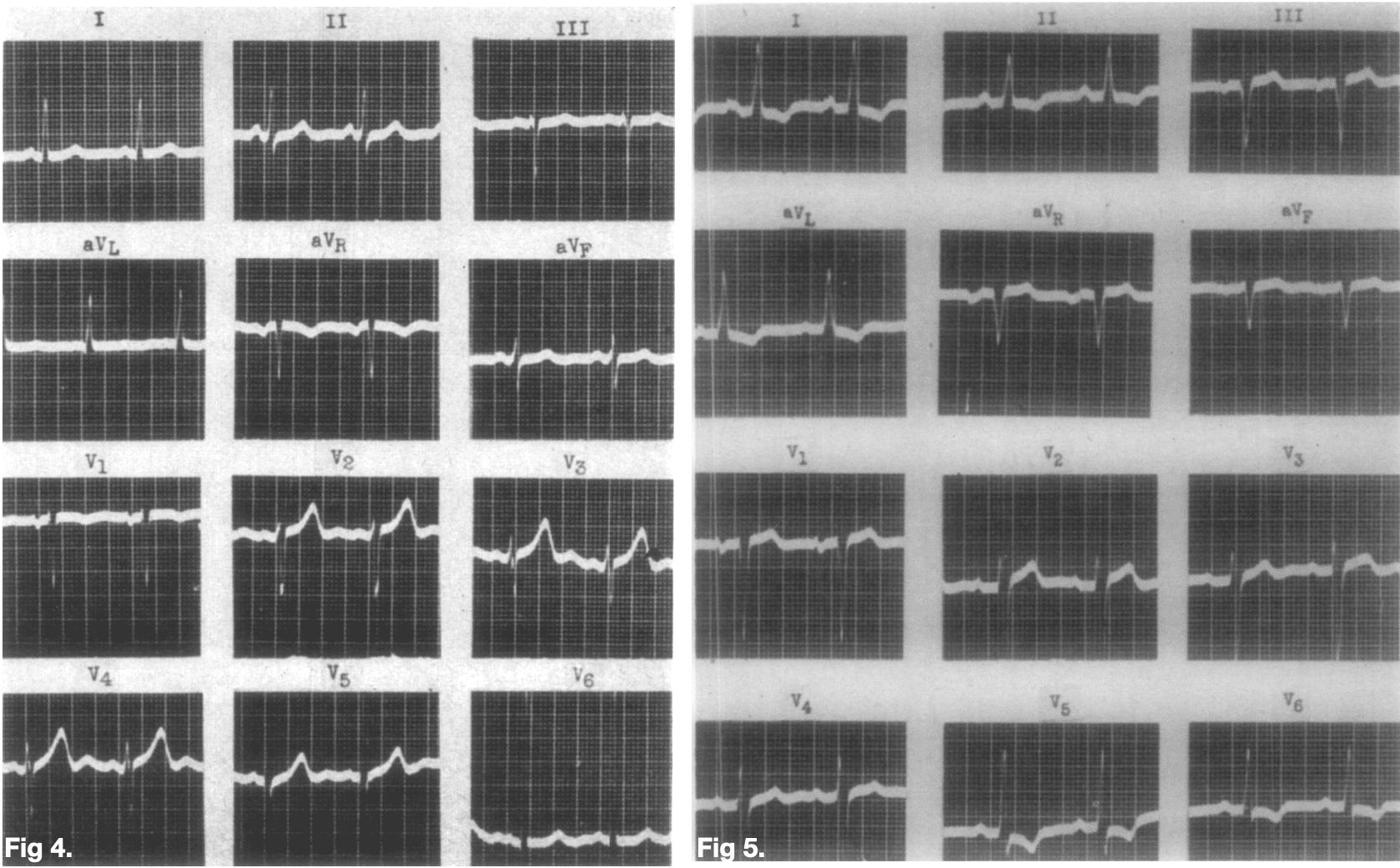
Figure 4: early LVH in a semihorizontal heart. The standard limb leads were largely unremarkable, but measured 44 mm.
Figure 5: more developed LVH with lateral ST-segment depression and asymmetric T-wave inversion—the classical secondary repolarisation or “strain” pattern.
Major Publications
- Sokolow M, Lyon TP. Criteria for the diagnosis of right ventricular hypertrophy using unipolar limb and precordial leads. Am J Med. 1947 Jul;3(1):125.
- Sokolow M, Lyon TP. The ventricular complex in left ventricular hypertrophy as obtained by unipolar precordial and limb leads. Am Heart J. 1949 Feb;37(2):161-86 [Sokolow-Lyon criteria]
- Sokolow M, Lyon TP. The ventricular complex in right ventricular hypertrophy as obtained by unipolar precordial and limb leads. Am Heart J. 1949 Aug;38(2):273-94.
- Sokolow M, Edgar AL. A study of the V leads in congenital heart disease; with particular reference to ventricular hypertrophy and its diagnostic value. Am Heart J. 1950 Aug;40(2):232-51.
- Sokolow M, Edgar AL. Blood Quinidine Concentrations as a Guide in the Treatment of Cardiac Arrhythmias. Circulation, 1950; 1(4): 576-592.
- Grubschmidt HA, Sokolow M. The reliability of high voltage of the QRS complex as a diagnostic sign of left ventricular hypertrophy in adults. Am Heart J. 1957 Nov;54(5):689-94
- Sokolow M, McIlroy MB. Clinical cardiology. 1977 (2e 1979 – 6e 1993)
- Perloff D, Sokolow M, Cowan R. The prognostic value of ambulatory blood pressures. JAMA. 1983 May 27;249(20):2792-8.
- Sokolow M. Ambulatory blood pressure. A personal historical account. Am J Hypertens. 1993 Jun;6(6 Pt 2):161S-165S.
References
Biography
- Maurice Sokolow, MD, pioneer in study of hypertension, dies. UCSF September 30, 2002
- Hall C. Dr. Maurice Sokolow – UCSF cardiologist. SFgate 2002
- Maurice Sokolow. Professor of Cardiology. University of California
Eponymous terms
- Romhilt DW, Estes EH Jr. A point-score system for the ECG diagnosis of left ventricular hypertrophy. Am Heart J. 1968 Jun;75(6):752-8.
- ArchiveGrid : E. Harvey Estes Jr. oral history interviews, 1990-2007 (oclc.org)
- Rautaharju P. History of electrocardiology: nine decades of ECG criteria for left ventricular hypertrophy–the contribution of Morice Sokolow and Thomas Lyon. Ann Noninvasive Electrocardiol. 2001 Oct;6(4):342.
- Peguero JG, Lo Presti S, Perez J, Issa O, Brenes JC, Tolentino A. Electrocardiographic Criteria for the Diagnosis of Left Ventricular Hypertrophy. J Am Coll Cardiol. 2017 Apr 4;69(13):1694-1703
- Estes EH, Zhang ZM, Li Y, Tereshchenko LG, Soliman EZ. Individual components of the Romhilt-Estes left ventricular hypertrophy score differ in their prediction of cardiovascular events: The Atherosclerosis Risk in Communities (ARIC) study. Am Heart J. 2015 Dec;170(6):1220-6.
- ECG in left ventricular hypertrophy (LVH): criteria and implications. ECG Waves
- Cadogan M. ECG criteria for left ventricular hypertrophy. LITFL
Eponym
the person behind the name
BSc, MD (UWA), MPH (UNSW). Emergency Physician. German translator and lover of medical history.
BA MA (Oxon) MBChB (Edin) FACEM FFSEM. Emergency physician, Sir Charles Gairdner Hospital. Passion for rugby, medical history, medical education, and asynchronous learning #FOAMed evangelist. Editor-in-Chief and Webmaster of Life in the Fast lane, LITFL | On Call 4e| Eponyms | Books | Horology |