![]()
Melioidosis a disease of surprises
Melioidosis is a fascinating disease, with a distinct geography, a wide range of clinical presentations and a complex pathogenesis. These factors all contribute to the public health challenge it presents in this region.
The clinical presentation of melioidosis varies from a rapidly fatal, septicaemic illness without pneumonia, to focal abscess-forming infection and asymptomatic exposure leading to seroconversion (1). The septicaemic illness is unusual because it can recur after several days or even weeks of apparently adequate intravenous antibiotic treatment. This feature of septicaemic melioidosis has prompted recommendations for follow-on eradication treatment for several months. Another unusual feature of septicaemic melioidosis is that it can occur a long time after the initial exposure (2, 3). This is thought to have its origins in a dormant subclinical infection. Other forms of severe infection include pneumonia that does not respond to conventional antibiotic therapy, and central nervous system infection which has a high mortality rate. Even in centres familiar with the infection, the mortality rate is high for these forms of melioidosis.
The subacute infections are usually focal and may affect almost any organ system. They can also act as a source for subsequent bacteraemia (4). Subclinical infection has been inferred from the incidental finding of positive melioidosis serology in apparently healthy adults. In some locations where overt infection is relatively common, seropositivity rates may be high. The lifelong risk of dormant infection progressing to late onset septicaemic disease is unknown, but presumed to be low. Little has been done to match melioidosis seroprevalance with exposure and subsequent infection. One notable exception is a recent prospective surveillance study from Taiwan (5).
From a public health point of view, melioidosis is a disease of mainly tropical locations, particularly in Southeast Asia and northern Australia (6). It has been recognised in many parts of the tropics and in a few subtropical locations. Cases occur sporadically throughout the endemic region, particularly during the tropical rainy season. People with occupational or recreational exposure to moist soil or surface water are at greatest risk. These include rice farmers, other agricultural workers, building site labourers, adventure travellers, soldiers and a variety of indigenous groups. A collection of co- morbidities (including diabetes, chronic renal failure, high alcohol consumption) are strongly associated with severe infection and poor outcomes. Very few true, point-source outbreaks of melioidosis have been recognised, and these have usually been associated with exposure to contaminated water (7-9).
Melioidosis is a bacterial infection caused by an oxidase positive, Gram negative bacillus known as Burkholderia pseudomallei. This species is easily grown on non-selective agar, is motile and almost universally resistant to Gentamicin and Colistin. These features can be used to arrive at a presumptive laboratory identification. Confirmation of its identity can be technically difficult due to a variety of closely related bacterial species including the low virulence, near-neighbour species such as Burkholderia thailandensis. We have found substrate utilisation panels unreliable for this purpose. Instead we use a collection of confirmatory tests including nucleic acid amplification, recombinase gene sequencing and bacterial cell wall fatty acid analysis (10, 11). Others have successfully used an in-house agglutinating antibody (12). B. pseudomallei colonies on agar media is said to be characteristic due to their wrinkled appearance (Figure 1). This feature should be used with caution because a proportion of isolates form smooth colonies and some can be overtly mucoid, similar to Pseudomonas aeruginosa (13).

Figure 1. Wrinkled colonies of B. pseudomallei on horse blood agar after five days of incubation, also showing pronounced haemolysis.
Resistance to aminoglycoside and cephalosporin antibiotics mean that these first-line intravenous agents are unsuitable for treatment in septicaemic infection. The antibiotic resistance pattern in the laboratory may therefore be an early indication of septicaemic melioidosis. The preferred antibiotics are Ceftazidime and Meropenem during the septicaemic phase of the disease, but these must be followed up with oral therapy; usually a combination of at least two agents from a list that includes cotrimoxazole, doxycycline and coamoxyclav (1). The quinolones have little place in melioidosis therapy though intracellular killing studies suggest that they may have an occasional role as an adjunct to another antibiotic with poorer intracellular penetration (2, 14).
A series of personal encounters with this disease illustrate some of the reasons why it continues to surprise physicians, pathologists and epidemiologists.
In Singapore during the 1990 there were reports of sudden deaths in Thai construction site workers, some of which were attributed to melioidosis (15). The severity of septicaemic melioidosis is such that it can cause death very shortly after exposure to contaminated soil or surface water. Clearly, a heavily populated location in the tropics such as Singapore can be expected to take a close interest in the public health implications, but it also raises questions about where else in the region sudden deaths from melioidosis might occur. The more typical, sporadic melioidosis cases in Singapore were confirmed by culture-based methods that led, inadvertently to an erroneous aetiological diagnosis in several cases (16). Careful repetition of the methods used highlighted the problems caused by over- reliance on substrate utilisation for B. pseudomallei identification (17). It was also in Singapore that features of the ecology of B. pseudomallei led me to investigate the interaction of this species with the free living amoeba, Acanthamoeba castellani. This was the experiment that allowed observation of bacterial adherence to the amoeba surface and the inference that the flagellum was necessary for internalisation in an amoebic vacuole (18, 19).
In the summer of 1996, colleagues in a Public Health Laboratory where I was working spoke of a case of melioidosis in a former World War II prisoner of war who had been imprisoned in Asia. His bacterial diagnosis had not been made immediately and thus laboratory staff had been exposed to a potential occupational infection hazard. This was an introduction to the prolonged delayed onset decades after initial exposure, and also to the risks inherent in working with unexpected, unfamiliar and potentially dangerous bacterial species. The current record for delayed onset stands at over 60 years (3).
Six months after arriving in Perth, I was alerted to a series of three cases of septicaemic melioidosis in the same remote community in northwestern Australia. A series of phone calls to the local public health office, regional and pathology branch lab indicated that there were likely to additional cases. As there had been three deaths already, we organised to fly in a small outbreak investigation team the following week. There was concern that the summer rains were due soon, making access to the affected community difficult and possibly increasing numbers of melioidosis cases. The team were able to fly in before the rains started, conducted a detailed environmental survey including soil and water sampling, case finding and preliminary control measures (7, 20). Preliminary data suggested that the drinking water supply had been contaminated, but it was only when detailed water engineering information was made available a year later that we recognised the likely upstream point source and confirmed it by further bacteriological investigation (21). These observations led to additional studies into Chlorine susceptibility of B.pseudomallei and persistence in acidic water at the low pH values prevalent in parts of WA (22, 23). One of the unanticipated benefits of these studies was an improved molecular biology capability. We built up molecular epidemiology methods with assistance from neighbouring centres (20), introduced rapid genotyping with added spin-offs for food microbiology (24, 25) and began using nucleic acid amplification techniques to identify bacteria (10).
Once we had an improved set of laboratory methods, we were in a stronger position to help our neighbours in the region. We joined in a series of field studies in Eastern Malaysia, exploring the upper reaches of the Sibu River in an attempt to map out the distribution of melioidosis. There have been cases of infection in forestry and construction workers quite far up river. However, there have been no cases reported from the remote communities of Barrio and Ba Kelalan further upstream where fragrant rice is grown by traditional methods (Figure 2a). Extensive soil sampling by our team in the latter community failed to recover any B pseudomallei from moist soil at optimal depth in rice paddies (Figure 2b). Further studies are under way into the ecology of Burkholderia species in Eastern Malaysia.
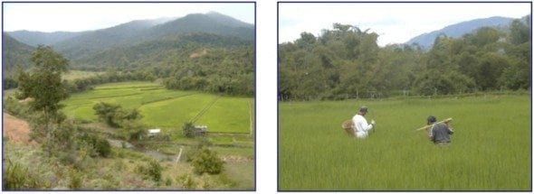
Figure 2a (left) highland rice fields at Ba Kelalan, Eastern Malaysia; and figure 2b (right) sampling the soil of rice fields using an augur.
The melioidosis outbreak in northeastern Brazil was another surprising development. Local investigation into a series of fatal febrile respiratory infections in a single family recovered a Gram negative bacillus from the blood cultures of one child. The infection was first thought to be plague, and the correct identification was not made before the child succumbed to the disease. After prolonged on-line discussion, we agreed to assist. A preliminary visit established a plan of action for the state Health Secretariat and a team was formed. During subsequent visits, members of our group travelled out to Brazil to train local people and develop laboratory capacity. Isolates were shipped back to Perth for detailed molecular analysis. The investigation team eventually published its findings, but not before there had been two further clusters of disease (9). Genotyping the collection of B. pseudomallei isolates showed that there was sufficient diversity to infer that the disease may have been present in Brazil for some time. By the relatively low discrimination ribotype method, the initial Brazil outbreak and the WA outbreak of 1997 appeared to be closely related. The possibility of a
historic link between these two outbreaks led to our observation that the origins of the disease may not necessarily lie in Southeast Asia or northern Australia (26). They could have been in the Americas, and have been followed by dissemination of infection through trade of agricultural stock such as domestic animals or crop plants. The proposals we have considered so far are goats, rubber and cassava.
We can assemble a short list of surprises from this series of snapshots;
- Epidemiology:
- Outbreaks of melioidosis do occur.
- Disease can be delayed onset or relapsing which may cause treatment failure.
- New locations continue to be identified in and at margins of endemic zone
- Bacteriology:
- B. pseudomallei can occur as Gram positive cocci under conditions of environmental stress.
- A mucoid variant phenotype has been observed that does not form wrinkled colonies.
- Identification difficulties have been experienced with substrate utilisation.
- Biodiversity & distribution of the Burkholderias cause interpretive difficulties with serology.
- Ecology:
- B. pseudomallei persists for longer periods in distilled water.
- It tolerates normally bactericidal Chlorine concentrations.
- Its size is optimal for inhalation and retention in the lungs when inhaled.
- A viable but non-culturable state has been inferred.
Some of these surprises have provided the challenge to spur further developments in the clinical laboratory:
- Molecular epidemiology:
- Techniques used for our B. pseudomallei genotyping include REPS-PCR, DNA macrorestriction, ribotyping, multi-locus sequence typing, variable number tandem repeat analysis and whole genome sequencing.
- Molecular diagnostics:
- Molecular diagnostic methods we have used for aetiolgical diagnosis of melioidosis nucleic acid amplification of the 16-23s spacer region of the genome, the LpxO and RecA loci.
- Water micro, other PHL methods:
- Drinking water treatment methods have been re-assessed. Bacterial particle size has been measured.
- Portable lab:
- Portable systems are under development, and have been used so far in the Kimberley, Brazil and will be employed in Sri Lanka. Emerging Infectious Diseases response capability has been expanded on this basis. Insights have been disseminated via the Australian PHLN, WHO-GOARN, other partners in the Indian Ocean rim, and through the UK Health Protection Agency.
- General diagnostic lab:
- Improved integration of molecular diagnostic and epidemiology methods has brought wider benefits to the clinical laboratory. Further developments in the application of molecular methods to blood cultures are now under way. These are expected to produce diagnostic benefits in our remote branch laboratories.
These developments only occurred because of an unusual level of collaboration at local, national and international level. A long list of friends and colleagues has contributed, as is evident in the reference list. We expect that this list will continue to grow, particularly now that we are using melioidosis to help us understand the role of the environment in the causation of infectious diseases (27).
References
- Inglis TJ, Rolim DB, Rodriguez JL. Clinical guideline for diagnosis and management of melioidosis. Rev Inst Med Trop Sao Paulo. 2006 Jan-Feb; 48(1):1-4. Epub 2006 Mar 9 [Reference].
- Inglis TJ, Golledge CL, Clair A, Harvey J. Case report: recovery from persistent septicemic melioidosis. Am J Trop Med Hyg. 2001 Jul; 65(1):76-82.
- Ngauy V, Lemeshev Y, Sadkowski L, Crawford G. Cutaneous melioidosis in a man who was taken as a prisoner of war by the Japanese during World War II. J Clin Microbiol. 2005 Feb; 43(2):970-2.
- Dhiensiri T, Eua-Ananta Y. Visceral abscess in melioidosis. J Med Assoc Thai. 1995 May; 78(5):225-31.
- Su HP, Yang HW, Chen YL, Ferng TL, Chou YL, Chung TC, Chen CH, Chiang CS, Kuan MM, Lin HH, Chen YS. Prevalence of melioidosis in the Er-Ren River Basin, Taiwan: implications for transmission. J Clin Microbiol. 2007 Aug; 45(8):2599-603. Epub 2007 Jun 27.
- Cheng AC, Currie BJ. Melioidosis: epidemiology, pathophysiology, and management. Clin Microbiol Rev. 2005 Apr; 18(2):383-416.
- Inglis TJ, Garrow SC, Adams C, Henderson M, Mayo M. Dry-season outbreak of melioidosis in Western Australia. Lancet. 1998 Nov 14; 352(9140):1600.
- Currie BJ, Mayo M, Anstey NM, Donohoe P, Haase A, Kemp DJ. A cluster of melioidosis cases from an endemic region is clonal and is linked to the water supply using molecular typing of Burkholderia pseudomallei isolates. Am J Trop Med Hyg. 2001 Sep; 65(3):177-9.
- Rolim DB, Vilar DC, Sousa AQ, Miralles IS, de Oliveira DC, Harnett G, O’Reilly L, Howard K, Sampson I, Inglis TJ. Melioidosis, northeastern Brazil. Emerg Infect Dis. 2005 Sep; 11(9):1458-60.
- Inglis TJ, Merritt A, Chidlow G, Aravena-Roman M, Harnett G. Comparison of diagnostic laboratory methods for identification of Burkholderia pseudomallei. J Clin Microbiol. 2005 May; 43(5):2201-6.
- Merritt A, Inglis TJ, Chidlow G, Harnett G. PCR-based identification of Burkholderia pseudomallei. Rev Inst Med Trop Sao Paulo. 2006 Sep-Oct; 48(5):239-44.
- Amornchai P, Chierakul W, Wuthiekanun V, Mahakhunkijcharoen Y, Phetsouvanh R, Currie BJ, Newton PN, van Vinh Chau N, Wongratanacheewin S, Day NP, Peacock SJ. Accuracy of Burkholderia pseudomallei identification using the API 20NE system and a latex agglutination test. J Clin Microbiol. 2007 Nov; 45(11):3774- 6. Epub 2007 Sep 5.
- Howard K, Inglis TJ. Novel selective medium for isolation of Burkholderia pseudomallei. J Clin Microbiol. 2003 Jul; 41(7):3312-6.
- Inglis TJ, Rodrigues F, Rigby P, Norton R, Currie BJ. Comparison of the susceptibilities of Burkholderia pseudomallei to meropenem and ceftazidime by conventional and intracellular methods. Antimicrob Agents Chemother. 2004 Aug; 48(8):2999-3005.
- Yap EH, Chan YC, Goh KT, Chao TC, Heng BH, Thong TW, Singh M, Jacob E. Pseudomonas pseudomallei and sudden unexplained death in Thai construction workers. Lancet. 1990 Aug 11; 336(8711):376-7.
- Ti TY, Tan WC, Chong AP, Lee EH. Nonfatal and fatal infections caused by Chromobacterium violaceum. Clin Infect Dis. 1993 Sep; 17(3):505-7.
- Inglis TJ, Chiang D, Lee GS, Chor-Kiang L. Potential misidentification of Burkholderia pseudomallei by API 20NE. Pathology. 1998 Feb; 30(1):62-4.
- Inglis TJ, Rigby P, Robertson TA, Dutton NS, Henderson M, Chang BJ. Interaction between Burkholderia pseudomallei and Acanthamoeba species results in coiling phagocytosis, endamebic bacterial survival, and escape. Infect Immun. 2000 Mar; 68(3):1681-6.
- Inglis TJ, Robertson T, Woods DE, Dutton N, Chang BJ. Flagellum-mediated adhesion by Burkholderia pseudomallei precedes invasion of Acanthamoeba astronyxis. Infect Immun. 2003 Apr; 71(4):2280-2.
- Inglis TJ, Garrow SC, Adams C, Henderson M, Mayo M, Currie BJ. Acute melioidosis outbreak in Western Australia. Epidemiol Infect. 1999 Dec; 123(3):437-43.
- Inglis TJ, Garrow SC, Henderson M, Clair A, Sampson J, O’Reilly L, Cameron B. Burkholderia pseudomallei traced to water treatment plant in Australia. Emerg Infect Dis. 2000 Jan-Feb; 6(1):56-9.
- Howard K, Inglis TJ. The effect of free chlorine on Burkholderia pseudomallei in potable water. Water Res. 2003 Nov; 37(18):4425-32.
- Howard K, Inglis TJ. Disinfection of Burkholderia pseudomallei in potable water. Water Res. 2005 Mar; 39(6):1085-92.
- Inglis TJ, O’Reilly L, Foster N, Clair A, Sampson J. Comparison of rapid, automated ribotyping and DNA macrorestriction analysis of Burkholderia pseudomallei. J Clin Microbiol. 2002 Sep; 40(9):3198-203.
- Inglis TJ, Clair A, Sampson J, O’Reilly L, Vandenberg S, Leighton K, Watson A. Real-time application of automated ribotyping and DNA macrorestriction analysis in the setting of a listeriosis outbreak. Epidemiol Infect. 2003 Aug; 131(1):637-45.
- Inglis TJ, Sagripanti JL. Environmental factors that affect the survival and persistence of Burkholderia pseudomallei. Appl Environ Microbiol. 2006 Nov; 72(11):6865-75. Epub 2006 Sep 15.
- Inglis TJ. Principia aetiologica: taking causality beyond Koch’s postulates. J Med Microbiol. 2007 Nov; 56(Pt 11):1419-22.
CLINICAL CASES
Tropical Travel Trouble
BA MA (Oxon) MBChB (Edin) FACEM FFSEM. Emergency physician, Sir Charles Gairdner Hospital. Passion for rugby; medical history; medical education; and asynchronous learning #FOAMed evangelist. Co-founder and CTO of Life in the Fast lane | On Call: Principles and Protocol 4e| Eponyms | Books |