![]()
Mirror Image Artefacts
Reverberation Artefact Refresh
An ultrasound machine assumes a single pulse of ultrasound enters the tissues, is reflected off a structure, and returns directly to the transducer for interpretation. When this does not occur ultrasound artefacts are created.
Not infrequently an ultrasound pulse encounters two parallel reflective surfaces lying perpendicular to its path. Some of the pulse becomes caught between the two surfaces, bouncing forwards and backwards before returning in increments, between each reverberation, to the transducer. This reverberation causes a repetitive artefact on the ultrasound image.
The appearance of the reverberation artefact depends on:
- The size of the two reflective surfaces
- The distance between the two reflective surfaces (long vs short path reverberation artefacts)
- How much ultrasound energy is lost – dissipated or attenuated, between each re-reflection.
Mirror image artefacts
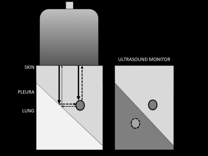
Mirror image artefact is one of the beam path artefacts. These occur when an ultrasound beam is not reflected directly back to the transducer after hitting a reflective surface, but rather takes an indirect return journey.
With mirror image artefact the interrogating ultrasound beam hits an angled, highly reflective surface, such a soft tissue / gas interface. It is reflected off into surrounding soft tissue (angle of incidence = angle of reflection). When it encounters a second reflective object it then begins its return journey to the transducer – along the same path.
The ultrasound machine cannot determine that the beam has taken an indirect return journey and a mirror image is created deep to the mirror surface. The appearance of the reflected image deeps on the smoothness, the angle, and the curvature of the mirror surface.
Two images are seen, the true image above the reflective surface, and the mirror image below.
How mirror image artefact is formed
An example of mirror image artefact
Echogenic benign cavernous haemangioma in the liver is seen above the diaphragm as well as below. The mirror artefact is larger than the true lesion because the mirror surface of the diaphragm is curved.
Mirror image artefact explained
Further Reading
- Long path reverberation artefact (A-lines in lung)
- Short path reverberation artefact (B-lines in lung; ringdown anywhere else!)
- Fatiguing short path reverberation artefact (Comet tails)
ULTRASOUND LIBRARY
Basics

An Emergency physician based in Perth, Western Australia. Professionally my passion lies in integrating advanced diagnostic and procedural ultrasound into clinical assessment and management of the undifferentiated patient. Sharing hard fought knowledge with innovative educational techniques to ensure knowledge translation and dissemination is my goal. Family, wild coastlines, native forests, and tinkering in the shed fills the rest of my contented time. | SonoCPD | Ultrasound library | Top 100 | @thesonocave |