


Morton neuroma
Morton neuroma is a painful compressive neuropathy of a common plantar digital nerve, classically affecting the third intermetatarsal space. The lesion is characterised by neural degeneration, epineural hyalinisation and perineural fibrosis. This produces a fusiform enlargement of the interdigital nerve distal to the deep transverse metatarsal ligament and before division into the digital branches.
The mechanism is multifactorial. Chronic mechanical irritation or compression of the interdigital nerve beneath the deep transverse metatarsal ligament is key. Contributing factors include the relatively large third-webspace nerve, mobility of the adjacent fourth ray, repetitive forefoot loading, tight or high-heeled footwear, and trauma.
Epidemiology
Morton neuroma predominantly affects middle-aged women. Di Caprio et al, found the female-to-male ratio was 4:1 and the mean age at surgery was about 50 years. The third intermetatarsal space accounted for approximately 66% of lesions, the second for 32% and the fourth for 2%. Bilateral disease was reported in about 21%, while multiple neuromas in the same foot were found to be uncommon.
Clinical presentation and diagnosis
Patients typically describe burning or shooting plantar forefoot pain between the metatarsal heads, commonly radiating into the adjacent toes. Numbness, tingling or an electric-shock sensation may occur. Symptoms are usually aggravated by walking, tight shoes or high heels and relieved by removing the shoe and massaging the foot.
Diagnosis is principally clinical. Examination aims to localise tenderness to the intermetatarsal space rather than directly beneath a metatarsal head. Compression of the forefoot while applying pressure to the affected interspace may reproduce pain and a palpable or audible click known as the Mulder sign or Mulder click.
How to elicit the Mulder sign: Compress the metatarsal heads together with one hand while applying firm plantar pressure to the symptomatic intermetatarsal space with the thumb of the other hand. Reproduction of the patient’s typical pain, with or without a palpable or audible click as the interdigital lesion displaces between the metatarsal heads, constitutes a positive test.
Plain radiographs are useful mainly to exclude other causes of metatarsalgia. Ultrasound or MRI may support the diagnosis when the clinical picture is uncertain or more than one webspace is suspected. However, asymptomatic interdigital nerve enlargement is common and imaging findings must correlate with symptoms. Ultrasound is preferred when imaging is required. A diagnostic local anaesthetic injection can provide additional confirmation.
Management
Initial treatment is usually non-operative, with modification of aggravating footwear and a plantar orthosis or metatarsal unloading pad intended to reduce pressure across the symptomatic interspace. Local corticosteroid injection may provide short-term relief. Complete resolution occurs in only a minority and repeated injections carry risks including plantar fat-pad atrophy and skin change.
Persistent, function-limiting symptoms despite conservative treatment can be managed surgically. Options include decompression with division of the deep transverse metatarsal ligament or neurectomy of the affected common digital nerve.
History
1835 – Filippo Civinini (1805-1844) published Su d’un nervoso gangliare rigonfiamento alla pianta del piede. He described a fusiform swelling of the common plantar digital nerve in the third intermetatarsal space during cadaveric dissection. His report was anatomical, without an associated clinical syndrome.
1845 – Lewis Durlacher surgeon and chiropodist to the Queen of England described a painful plantar nerve syndrome in A Treatise on Corns, Bunions, the Diseases of Nails, and the General Management of the Feet. Durlacher provided an early clinical description of plantar neuralgia but did not name the condition.
Another form of neuralgic affection occasionally attacks the plantar nerve on the sole of the foot, between the third and fourth metatarsal bones, but nearest to the third, and close to the articulation with the phalanx. The spot where the pain is experienced can at all times be exactly covered by the finger. The pain, which cannot be produced by the mere pressure of the finger, becomes very severe whilst walking, or whenever the foot is put to the ground.
Durlacher 1845: 52
1876 – Thomas George Morton (1835-1903) published A peculiar and painful affection of the fourth metatarso-phalangeal articulation, a condition he claimed that had not previously been described. Morton described 16 cases of a distinctive painful syndrome centred clinically on the fourth metatarsophalangeal region, with severe paroxysmal pain provoked by shoe pressure.
Although the paper title emphasised the joint, Morton suspected a neural lesion “either neuroma or some nerve hypertrophy”. He proposed that the mobile fifth metatarsal could compress the fourth metatarsophalangeal region and adjacent plantar nerve branches..
1880 – In Painful affection of the foot, Morton expanded the clinical description. He noted that the fourth MTP joint was most often involved, but he had also seen cases affecting the third. He described mild and severe forms and recurrent neuralgic pain. He treated severe cases by excision of the of the painful joint and surrounding tissues.
1891 – American orthopaedic surgeon Edward Hickling Bradford (1848-1926) published Metatarsal neuralgia, or Morton’s affection of the foot. An early example of eponymous attribution.
The title “Morton’s Affection” could properly be used, as the affection was first thoroughly described by Dr. Thomas G. Morton, of Philadelphia; but a personal name is, as a rule, to be avoided, if possible, in defining a disease, and it is for this reason that the term “metatarsal neuralgia,” has been preferred. The disease consists of a form of neuralgia of the foot, quite clearly marked in its symptoms and often exceedingly distressing. The affection is not an uncommon one, but has hitherto attracted but little attention.
Bradford 1891
1893 – Morton’s son, Thomas Story Kirkbride Morton, published Metatarsalgia (Morton’s Painful Affection of the Foot) consolidating the neural explanation. He described the condition as a painful affection of the plantar digital nerves produced by compression or pinching of the plantar digital nerves. His operations remained directed at the fourth MTP joint and he acknowledged that no affected nerves had yet been excised for microscopic examination.
1893 – Albert E. Hoadley (1847–1900) reported Six cases of metatarsalgia. He challenged the Mortons’ proposed joint mechanism and argued that the digital nerve itself was the source of pain. In one patient he resected the nerve alone and found a small red fusiform enlargement which he regarded as a neuroma, with complete and lasting relief.
It being the digital nerve that is involved, the articulation… [has] but little to do with the painful affection.
1896 – Thomas George Morton revisited the condition using the newly introduced X-rays to the Diagnosis of Morton’s Painful Affection of the Foot, or Metatarsalgia. He described it as a painful affection of the plantar digital nerves caused by compression or pinching around the fourth and fifth MTP articulations.
Morton wrote that after twenty years he had found no reason “to modify or change my views as to its pathology.” He restated his original nerve-compression-with-joint-mechanics model. The X-rays were intended to demonstrate the osseous relationships and toe distortion that he believed contributed to the syndrome.
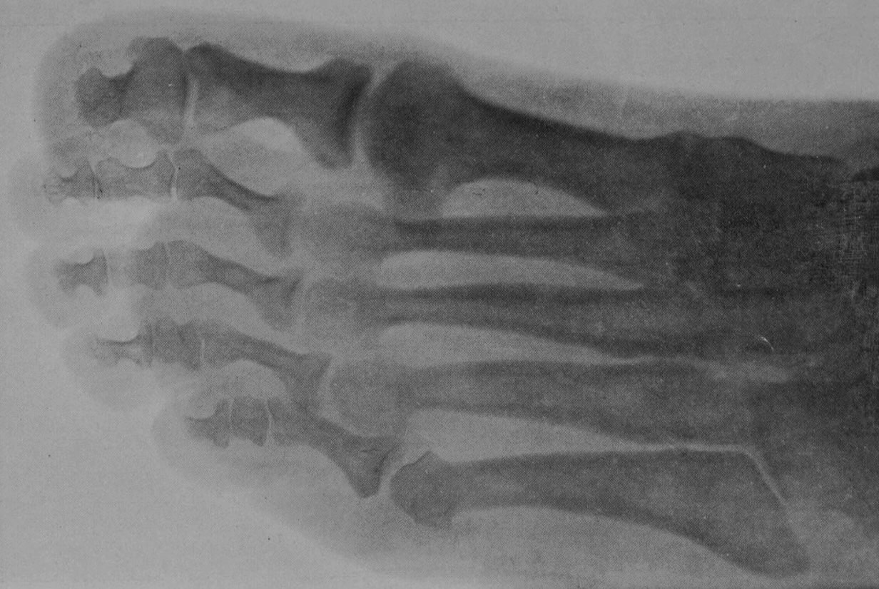
1927 – Dudley Joy Morton later cited Thomas George Morton’s 1896 radiographic paper in his own studies of forefoot morphology, providing an unusual link between the two unrelated “Mortons” of foot eponymy.
1940– L. O. Betts described Morton’s metatarsalgia as a neuritis of the fourth digital nerve with a pronounced neuroma in all cases. This helped formalise the neural interpretation of the syndrome.
1947 – Bickel and Dockerty published Plantar neuromas, Morton’s toe which helped associate the eponymous term with a pathological nerve lesion. They resurrected Hoadley’s contribution, noting that his findings had remained largely unknown for almost five decades, and credited Betts with re-establishing the nerve lesion and operative treatment.
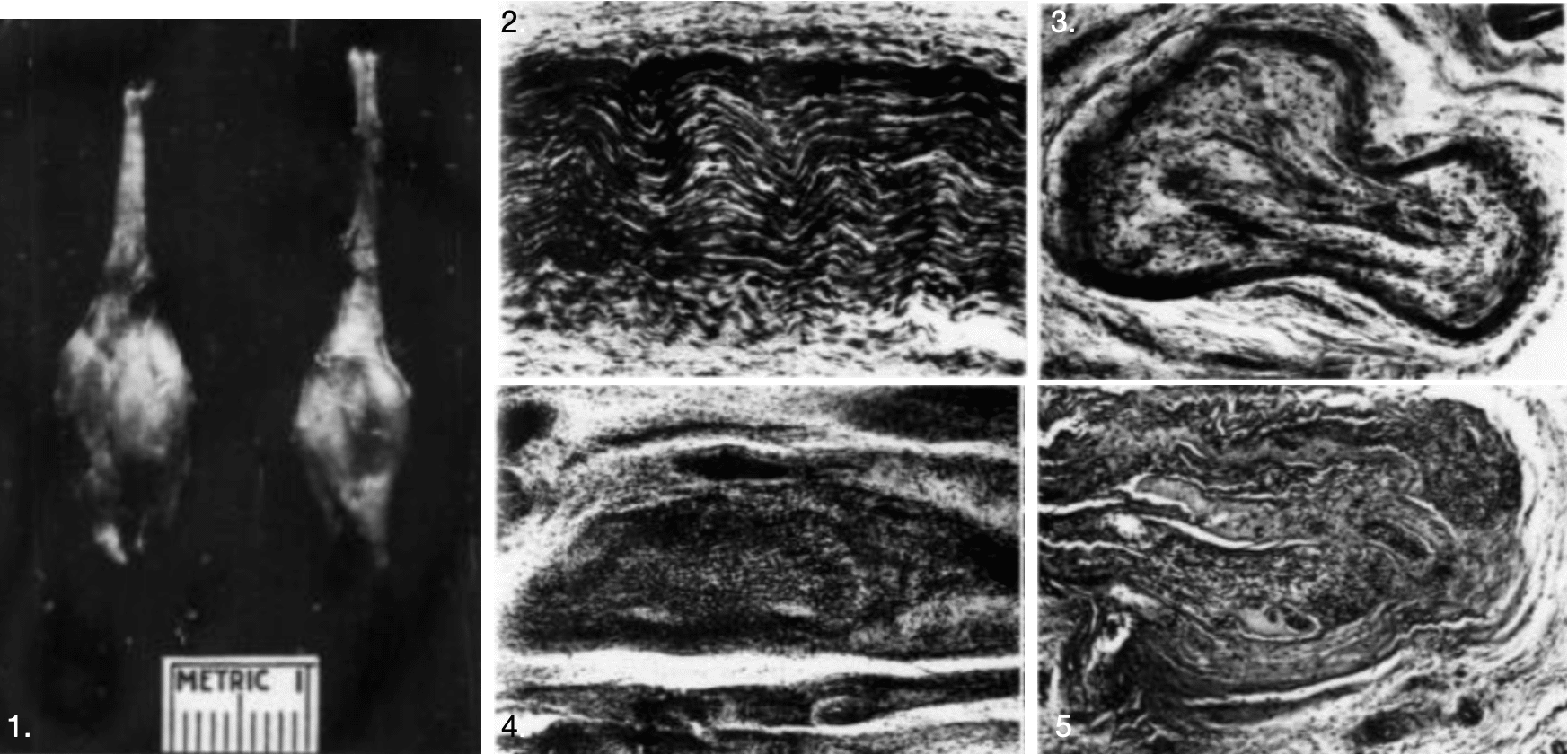
The pair provided gross and histological study of the excised lesions. They concluded that the “Morton’s toe” or “Morton’s metatarsalgia” had a pathological basis in tumefactive perineural fibrosis, with degenerative and proliferative nerve changes and neural/perineural oedema.
1951 – Jacob D. Mulder published The causative mechanism in morton’s metatarsalgia in which he reported the operative findings in 12 cases and discussed the mechanism of digital nerve compression. He also described a clinical manoeuvre for Morton metatarsalgia. He found that compression of the metatarsal heads could produce a characteristic painful click, the Mulder sign or Mulder click.
2005 – Larson et al argued that Morton neuroma is a misleading name. It is neither the precise lesion Morton originally demonstrated nor a true neoplasm. They favoured descriptive terminology such as compression of the interdigital nerve to the third web space or compression of the common plantar digital nerve.
2006 – Pasero and Marson emphasised Civinini’s 1835 anatomical priority and supported Civinini–Morton as more appropriate eponymous terminology. They noted that Civinini’s description of a fusiform nerve enlargement in the third intermetatarsal space closely corresponds to what is now recognised as the anatomical substrate of the modern lesion.
2018 – Di Caprio et al describe a fusiform interdigital nerve lesion, usually in the third intermetatarsal space, with neural degeneration, epineural hyalinisation and perineural fibrosis. Not a true neoplastic neuroma.
Eponymythology
Morton neuroma is an historically imprecise eponym. Filippo Civinini has priority for the anatomical lesion, describing a fusiform enlargement of a plantar digital nerve in 1835. Lewis Durlacher provided an early clinical description of plantar neuralgia in 1845. Thomas George Morton’s provided the detailed clinical description that established and popularised the painful forefoot syndrome subsequently associated with his name.
The concept changed substantially after Morton. His original syndrome was centred on painful compression around the fourth metatarsophalangeal articulation, albeit with suspected nerve involvement. Later authors progressively shifted the pathological focus from the articulation, to plantar digital nerve compression, and finally to a demonstrable fusiform nerve lesion with perineural fibrosis. The modern term neuroma is imperfect. The lesion is not a true neoplasm, but a chronic compressive neuropathy with degenerative and fibrotic change.
The name is further complicated by the Mortons. TG Morton described the original clinical syndrome, his son TSK Morton developed the nerve-compression explanation, and the unrelated DJ Morton later generated a separate family of foot eponyms.
The most confusing part is the Morton toe. Initially Morton toe referred to Thomas George Morton’s painful forefoot syndrome. Bickel and Dockerty used the term for plantar neuromas in 1947. However we now refer to the Morton toe based on Dudley Joy Morton’s short-first-metatarsal morphology. The same eponymous phrase therefore migrated from one Morton, and one disorder, to another.
Associated Persons
- Filippo Civinini (1805-1844)
- Lewis Durlacher (1792-1864)
- Thomas George Morton (1835-1903)
- Edward Hickling Bradford (1848-1926)
- Thomas Story Kirkbride Morton (1865-1930)
- Albert E. Hoadley (1847–1900)
- Jacob D. Mulder (1901–1965)
Alternative names
- Morton metatarsalgia, Morton neuralgia, Morton’s painful affection of the foot
- Interdigital neuroma, intermetatarsal neuroma, interdigital nerve compression
- Civinini–Morton neuroma.
References
Historical references
- Civinini F. Su d’un nervoso gangliare rigonfiamento alla pianta del piede. Lettera anatomica al Dr. Salomone Lampronti. Pistoia, Tip. Bracali, 1835
- Durlacher L. A treatise on corns, bunions, the diseases of nails, and the general management of feet. 1845: 52
- Morton TG. A peculiar and painful affection of the fourth metatarso-phalangeal articulation. American Journal of the Medical Sciences. 1876; 71: 37–45.
- Bradford EH. Metatarsal neuralgia, or Morton’s affection of the foot. Boston Medical and Surgical Journal 1891; 125: 52-55
- Morton TSK. VI. Metatarsalgia (Morton’s Painful Affection of the Foot), with an Account of Six Cases Cured by Operation. Ann Surg. 1893 Jun;17(6):680-99.
- Hoadley AE. Six cases of metatarsalgia. Chicago Medical Recorder 1893; 5: 32-37
- Morton TG. The Application of the X-rays to the Diagnosis of Morton’s Painful Affection of the Foot, or Metatarsalgia. International Medical Magazine 1896; 5(5): 322-324
- Jones R. Plantar neuritis. Liverpool medico-chirurgical journal. 1897; 17:
- Betts LO. Morton’s metatarsalgia: neuritis of the fourth digital nerve. Medical Journal of Australia 1940; 1(15): 514
- Bickel WH, Dockerty MB. Plantar neuromas, Morton’s toe. Surg Gynecol Obstet. 1947 Jan;84(1):111-6
- Mulder JD. The causative mechanism in morton’s metatarsalgia. J Bone Joint Surg Br. 1951 Feb;33-B(1):94-5.
Eponymous term review
- Larson EE, Barrett SL, Battiston B, Maloney CT Jr, Dellon AL. Accurate nomenclature for forefoot nerve entrapment. A historical perspective. Journal of the American Podiatric Medical Association 2005; 95(3): 298-306.
- G. Pasero, P. Marson. Filippo Civinini (1805-1844) and the discovery of plantar neuroma. Reumatismo 2006; 58(4): 319-22
- Di Caprio F, Meringolo R, Shehab Eddine M, Ponziani L. Morton’s interdigital neuroma of the foot. Foot and Ankle Surgery. 2018;24(2):92-98.
eponymictionary
the names behind the name

BA MA (Oxon) MBChB (Edin) FACEM FFSEM. Emergency physician, Sir Charles Gairdner Hospital. Passion for rugby, medical history, medical education, and asynchronous learning #FOAMed evangelist. Editor-in-Chief and Webmaster of Life in the Fast lane, LITFL | On Call 4e| Eponyms | Books | Horology |