Osteopoikilosis
Description
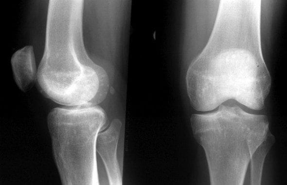
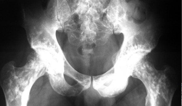
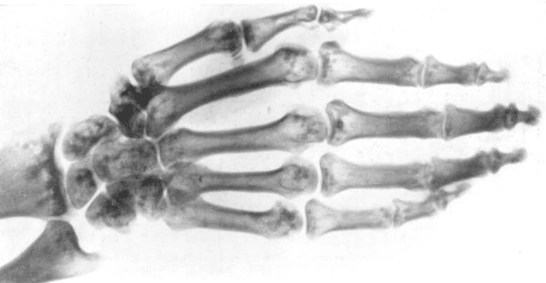
Osteopoikilosis is a autosomal dominant sclerosing bony dysplasia characterized by multiple benign benign sclerotic bone lesions (enostoses) that tend to localize in periarticular osseous regions
From the greek ποικίλος (poikilos) = spotted


History of osteopoikilosis
1915 – Albers-Schönberg described the case of a 22 year old male in whom roentgen examination revealed numerous minute lesions in all bones except the skull, spine, scapula, patella and clavicle. Lesions were located chiefly in the epiphyses and diaphyses and appeared as minute oval areas of condensation, usually with the long axis in the direction of the long axis of the bone. Their size varied from that of the head of a pin to that of a pea. They were sometimes so numerous as to make the bone look spotted in the roentgenogram.
Die in folgendem beschriebene eigentümliche Anomalie des Skelettes wurde zufällig bei der Röngenuntersuchung eines gesunden Soldaten gefunden. Meines Wissens ist ein ähnlicher oder gleicher Fall bisher nicht bekannt geworden.
Soldat K., 22 Jahre alt, im Zivilberuf Emaillebrenner.
Während des Krieges hat er bis zum heutigen Tage alle Strapazen gut überstehen können. Zur zeit ist er ins Lazarett geschickt wegen rechtsseitiger Schulterschmerzen, die so stark sind, daß er den Arm nicht ordentlich heben kann.
Bei der Röntgenuntersuchung des Schultergelenks und Fußgelenks ergab sich kein pathologischer Befund, dagegen fiel sofort folgende Skeletteigentümlichkeit auf:
Verteilt über die gesamten Fußknochen, mit Einschluß des unteren Teiles von Tibia und Fibula, finden sich etwa linsengroße Verdichtungsherde, ähnlich den bekannten Kompaktainseln. Diese Flecke stehen mit ihrer Längsachse stets in der Längsachse des betreffenden Skelett teiles.
Es handelt sich wohl um eine belanglose Erscheinung, die jedenfalls niemals klinische Bedeutung gewonnen zu haben scheint. Eine Brüchigkeit der Knochen liegt trotz des vermehrten Kompaktasubstrates nicht vor
The particular anomaly of the skeleton described here was discovered incidentally during radiological examination of a healthy soldier. To my knowledge there is no description of a similar case to date.
Soldier K, 22 years old, metalworker in civil service.
During the war he has thus far coped well with all efforts. He has been sent to the lazaret (hospital) because of right-sided shoulder pains, which are so strong, that he cannot lift his arm properly.
X-ray examination of the shoulder and ankle joints revealed no pathological findings, however the following skeletal particularity was immediately apparent:
Spread over the entire tarsal bones, including the lower parts of Tibia and Fibula, there are lenticular clusters of densification, similar to Enostoses. These spots are arranged such that their long axis follows the long axis of the respective bone in which they are found.
This must be an inconsequential appearance, at least one that has never merited clinical attention, it would seem. Despite the increased amount of compact substrate, there is no heightened fragility of the bones
1916 – René Ledoux-Lebard, Chabaneix and Dessane published on a further case, and coined the term l’ostéo-poecilie (osteopoikilosis).
Une radiographie du genou faite chez un blessé en vue d’une recherche de minimes fragments métalliques nous a révélé , il y a quelque temps déjà , un aspect très curieux du squelette de cette articulation …
Il s’agit de petits ilots sombres, ovalaires ou arrondis, ayant de 1 à 8mm…et qui tranchent nettement sur la teinte de l’os environnant…
Nous avons signalé que ces taches faisaient défaut dans le squelette de la tête ainsi que dans les vertèbres proprement dites, mais nous en constatons quelque unes dans le sacrum…
Elles sont tout particulièrement abondantes dans la tête de l’humérus et du fémur, le pourtour de la cavité cotyloïde, les condyles fémoraux et l’extrémité supérieure du tibia.
Nous n’avons pu constater chez notre sujet aucun symptôme clinique anormal. Sa santé générale semble parfaite.
Seul un diagnostic radiologique semble donc possible et nous proposons pour cet aspect osseux nouveau et si curieux la désignation d’ostéo-poecilie (ποικίλος : varié, moucheté) qui a l’avantage de ne rien préjuger à sa nature et à son origine
An X-ray of a knee performed on a wounded (soldier) to search for minimal metallic fragments has revealed to us, some time ago, a most curious aspect of the skeleton of this joint.
There are small, dark islets, oval or round, measuring 1 to 8 mm… demarcated clearly on the bone which they lie on…
We have signalled that these spots are not present in the cranium or the vertebrae, though we have noticed some in the sacrum…
They are particularly abundant in the heads of the humerus and femur, around the acetabulum, the femoral condyles and the superior extremity of the tibia.
We have not been able to elicit any abnormal clinical symptom in our subject. His general health appears perfect.
It would appear that only a radiological diagnosis is possible, and we propose, for this new and curious appearance of the bone, the designation of osteopoikilosis (ποικίλος : varied or speckled), which has the advantage of not presuming anything with regards to its nature.

Ledoux-Lebard et al. 1916-1917
At time of publication, the French and Germans were not on speaking terms due to the outbreak of the great war. Neither Schönberg nor Ledoux-Lebard et al. knew of the others’ case. In more peaceful times, the latter were kind enough to tip their hat to Schönberg, with an endnote in their publication
Note ajoutée à la correction:
Venant d’avoir l’occasion de parcourir les fascicules de Fortschritte auf dem Gebiete der Roentgenstrahlen parus depuis la guerre, nous avons pu constater dans l’un d’eux la publication par Albers-Schönberg d’un cas absolument comparable au nôtre. Pour être rare, le cas que nous décrivons n’est donc pas isolé et il s’agit bien là non d’une simple curiosité casuistique mais d’un processus nouveau, radiologiquement détectable, ce qui en réhausse considérablement l’intérèt.
Correction note:
Having had the chance to peruse the booklets of Fortschritte auf dem Gebiete der Roentgenstrahlen, which have appeared following the war, we have noted the publication by Albers-Schönberg of a case absolutely comparable to ours. Rare as it may be, the case we have presented is therefore not isolated and it is not a simple incidental curiosity, but rather a new process, radiologically detectable, which raises the interest in it considerably.
…the name given it by Ledoux-Lebard, Chabaneix and Dessane…is a good one, inasmuch as it is neutral as to the pathogenesis of the disease and the causes of the osseous changes involved, which still remain to be elucidated. The roentgenologic picture of osteopoikilosis is characterised by the presence of multiple round or somewhat oblong, sharply circumscribed but sometimes confluent, opaque spots, very much resembling ordinary osseous nuclei. They vary in size from a few millimetres in diameter to a couple of centimetres in length, and are localised chiefly to the spongiosa of the epiphyses and metaphyses of the long tubular bones, being usually massed most densely close to the articulations.
Petersen Fr. G 1936

Petersen Fr. G 1936
Associated Persons
- Heinrich Albers-Schönberg (1865 – 1921)
- René Ledoux-Lebard (1879–1948)
Alternative names
- ostéo-poecilie, ostéopoecilie
References
- Albers-Schönberg H. Eine seltene, bisher nicht bekannte Strukturanomalie des Skelettes. Fortschritte auf dem Gebiete der Röntgenstrahlen. 1915; 23: 174-175
- Ledoux-Lebard R, Chabaneix et Dessane. L’ostéopoécilie, forme nouvelle d’ostéite condensante généralisée sans symptomes cliniques. Journal de radiologie et d’électrologie. 1916-1917; 2: 133–134
- Petersen Fr. G. A Case of Osteopoikilosis. Acta Radiologica, 1936; 17: 388-396
[cite]
eponymictionary
the names behind the name
Resident medical officer in emergency medicine MB ChB (Uni. Dundee) MRCS Ed. Avid traveller, yoga teacher, polylinguist with a passion for discovering cultures.


Interesting article about a little-known condition