![]()
Rigler notch sign
The Rigler Notch Sign refers to a notched or umbilicated indentation along the border of a pulmonary mass, seen on radiographic imaging. It is traditionally interpreted as suggestive of malignancy, particularly bronchial carcinoma, though it can occur in benign conditions such as granulomatous disease or bronchial adenoma. The sign is most reliably observed on body-section roentgenograms and CT imaging.
Description
- Location: Typically along the medial or inferior margin of a spheroidal lung mass.
- Appearance: A sharp, focal indentation or groove on an otherwise smooth mass border.
- Interpretation: May represent a feeding vessel, retracted bronchus, or vascular hilus-like configuration within the tumour mass.
- Sensitivity/Specificity: While potentially indicative of malignancy, it is not pathognomonic, especially in small lesions.
Associated Features
- Pleural Tail Sign (“rabbit ears”); linear opacities extending to pleura, also described by Rigler (1965), often overlaps in interpretation.
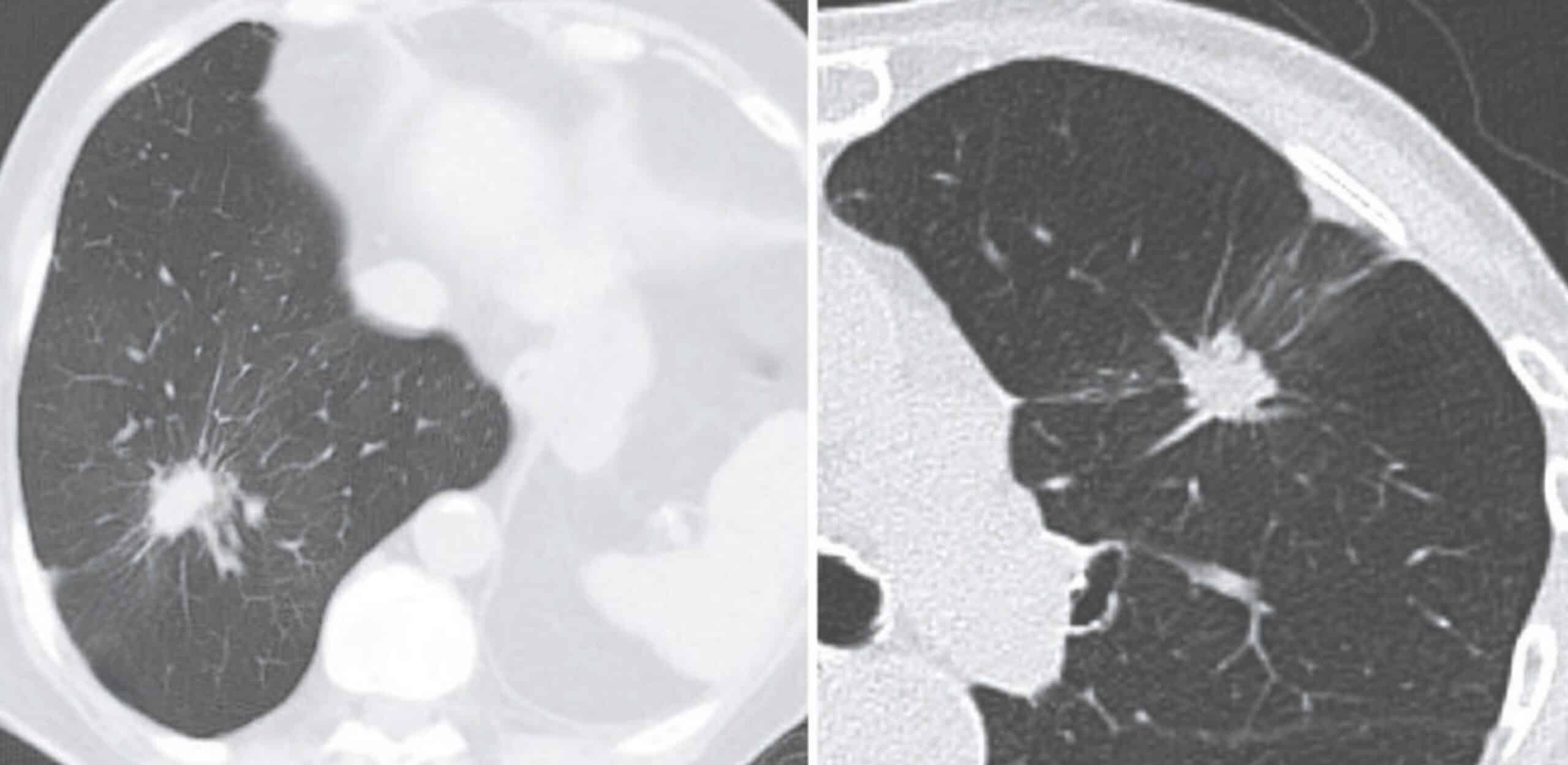
- “Navel-like retraction” seen on CT, associated with tumors overgrowing adjacent vessels.
- Positive bronchus sign may coexist, where a bronchus leads into but terminates within a mass.
Clinical Utility
- Use: Suggestive sign in radiographic assessment of solitary pulmonary nodules.
- Best demonstrated: On CT chest with contrast, especially thin-slice axial reconstructions.
- Limitations: Present in benign lesions, not diagnostic on its own.
History of the Rigler notch sign
1955 – Leo George Rigler (1896-1979) and E. Robert Heitzman (1927-2020) published findings of their blinded review of 132 lung films with spheroidal lung nodules. The study reviewed three radiological signs of lung nodules: calcification, cavitation and notching. The authors noted that calcification was associated with benign lesions and cavitation was indicative of malignancy. The authors describe a notch sign for the first time.
A new roentgen sign, the notching or umbilication of a border of a spheroidal nodule is described. It is best demonstrated in body-section roentgenograms. When definite, it is highly indicative of malignancy, either primary or metastatic. Wen equivocal, especially in very small lesions, it is of little value. The absence of this sign is of no significance.
Rigler and Heitzman, Radiology 1955
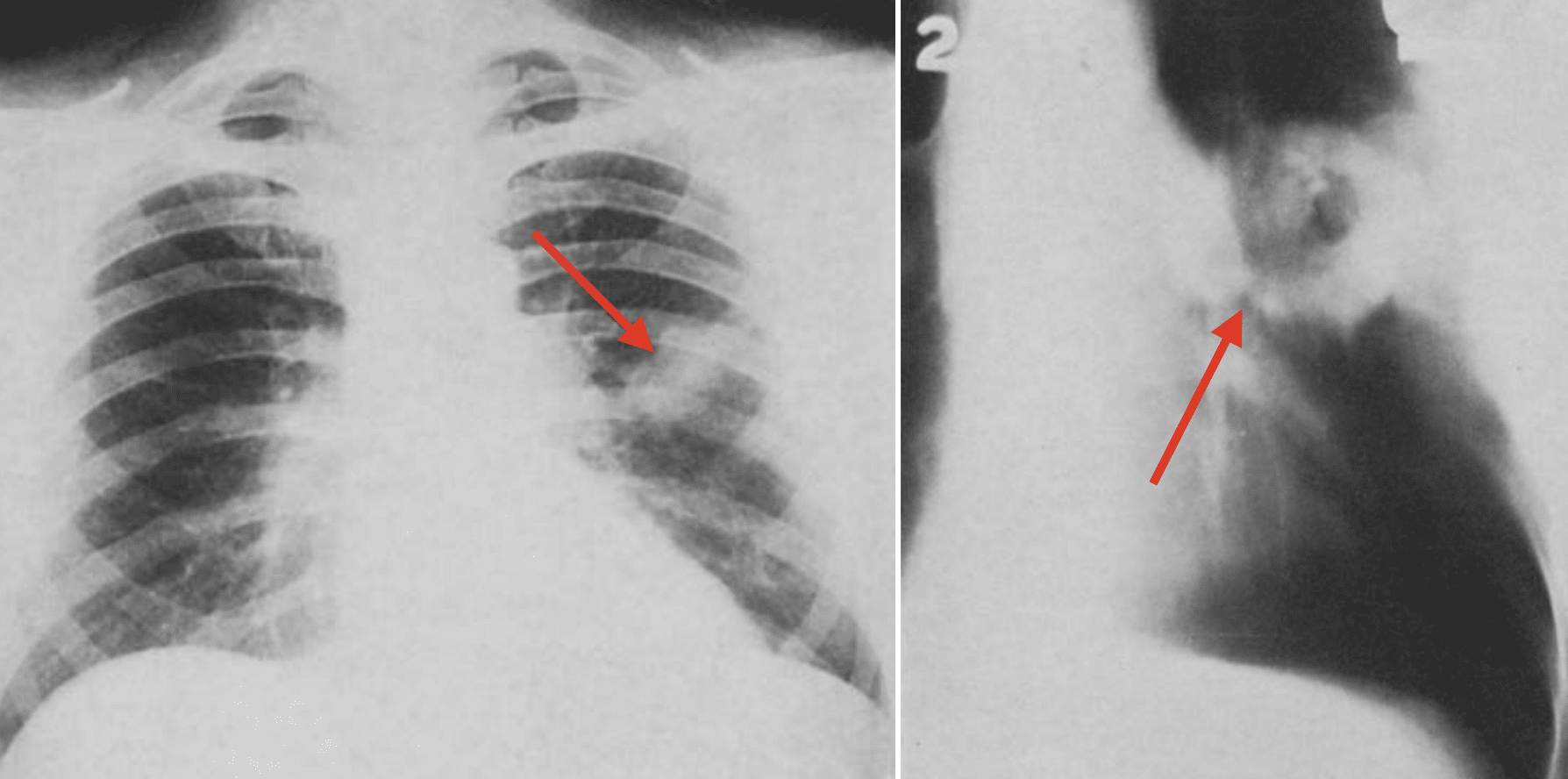
Fig. 5. Notch sign in a conventional roentgenogram. Squamous-cell carcinoma showing nodular density with notch sign on superior border (arrow). Radiology 1955
Rigler and Heitzman describe the notches pathologically and histologically.
In many histologic section of this small lesion shows an area of blood vessels surrounded by alveoli which is drawn into one side of the tumour. It is possible that the tumour has grown around this stalk, thus resembling the hilus of an organ.
Rigler and Heitzman, Radiology 1955
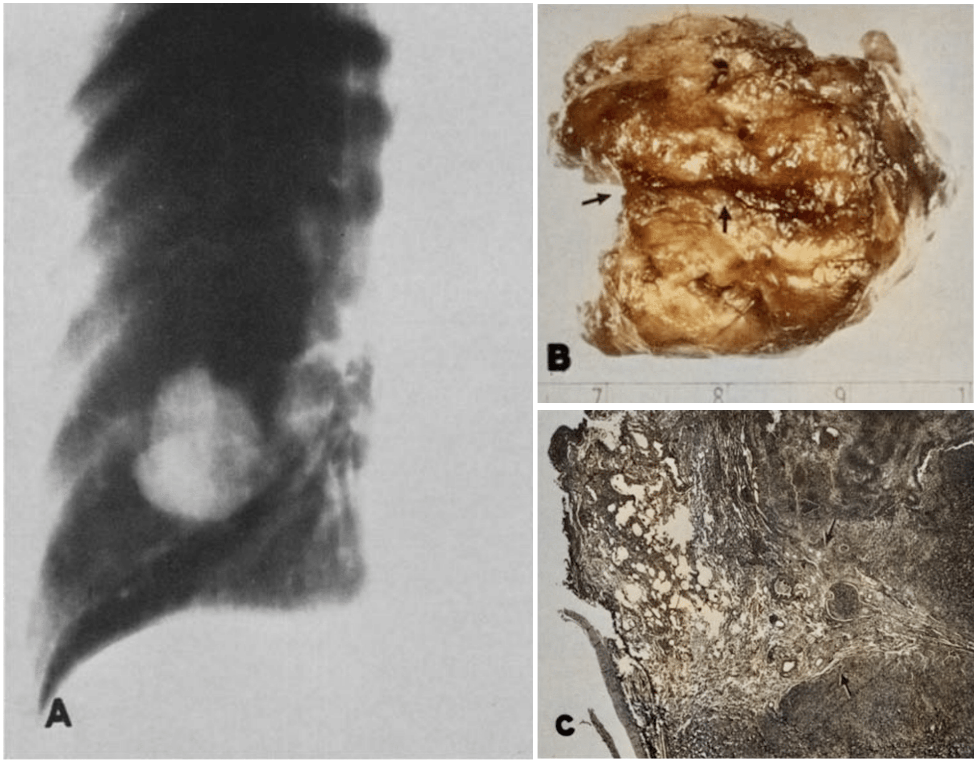
Fig. 8. Notch sign in a hypernephroma metastasis. A. Planigram showing large nodule in lung with marked indentation on lateral border, fairly characteristic of notch sign. B. Surgical specimen showing a deep groove (arrows) in the tumor corresponding to the notch. C. Microscopic section showing alveoli and blood vessels extending into the margin of the tumor. Radiology 1955
1965 – Rigler presents further radiological signs of bronchioloalveolar carcinoma (BAC) including the air bronchogram and pleural tail sign, building on the diagnostic value of peripheral contour irregularities.
…linear shadow running through the substance of the nodule, sometimes connecting it with the pleura, but not always, which appears to be a necrotic area within the tumor which has become dense and produced a puckering of the pleura.” For want of better descriptive term, this finding (present in 57 per cent of the patients in our series) has been termed tile “rabbit-ear” or “tail” sign.
Rigler 1965
pleural surface without interruption, and forming a wedgeshaped protrusion on the visceral pleura
Right: The presence of two lines running from the lesion to
the visceral pleura (or even more) is called rabbit ears. Yudin 2023
Associated Persons
- Leo George Rigler (1896-1979)
- E. Robert Heitzman (1927-2020)
Alternative names
- Notch Sign of Malignancy
References
Historical references
- Rigler LG, Heitzman ER. Planigraphy in the differential diagnosis of the pulmonary nodule, with particular reference to the notch sign of malignancy. Radiology. 1955 Nov;65(5): 692-702.
- Rigler LG. A roentgen study of the evolution of carcinoma of the lung. J Thorac Surg. 1957 Sep; 34(3): 283-297
Eponymous term review
- Shapiro R, Wilson GL, Yesner R, Shuman H. A useful roentgen sign in the diagnosis of localized bronchioloalveolar carcinoma. Am J Roentgenol Radium Ther Nucl Med. 1972 Mar;114(3):516-24.
- Eisenberg RL. Clinical Imaging: An Atlas of Differential Diagnosis [Internet]. Wolters Kluwer Health/Lippincott Williams & Wilkins; 2010
- Yudin A. Positive Bronchus Sign and Navel-Like Retraction, Rigler Notch Sign, Rigler Sign. In: Metaphorical Signs in Computed Tomography of Chest and Abdomen. 2023
- Gaillard F. Rigler notch sign (lungs). Radiopaedia.
eponymictionary
the names behind the name

Third year M.D. student at the University of Notre Dame Fremantle. Passionate about emergency and retrieval medicine, rural practice and clinical research
BA MA (Oxon) MBChB (Edin) FACEM FFSEM. Emergency physician, Sir Charles Gairdner Hospital. Passion for rugby; medical history; medical education; and asynchronous learning #FOAMed evangelist. Co-founder and CTO of Life in the Fast lane | On Call: Principles and Protocol 4e| Eponyms | Books |