


Rolando fracture
Rolando fracture is a complete intra-articular fracture of the base of the first metacarpal, classically dividing the base into three major components producing a Y- or T-shaped configuration.
The eponymous term is now commonly extended to intra-articular first-metacarpal base fractures with three or more fragments, although some authors distinguish severely comminuted fractures from the classic three-part Rolando pattern.
Rolando fractures are unstable injuries caused predominantly by axial loading of the thumb. Compared with the two-part Bennett fracture-dislocation, increasing articular fragmentation makes reduction and stable fixation more difficult and contributes to a less predictable functional outcome.
Mechanism and anatomy
Rolando fractures usually follow axial compression through a flexed or adducted thumb, as with a fall onto the radial side of the hand or impact through a clenched fist. Rolando recorded both mechanisms in his original three cases.
In the classic injury, a longitudinal intra-articular fracture line divides the metacarpal base into dorsal and volar articular fragments, while a transverse component separates these from the metacarpal shaft. The resulting Y- or T-shaped fracture therefore involves the entire articular surface rather than the single marginal fragment characteristic of a Bennett fracture. More severe injuries may produce multiple small articular fragments and central impaction.
Clinical features
Presentation resembles other first-metacarpal base fractures, with pain, swelling and tenderness over the thumb CMC joint, painful restriction of movement and reduced pinch or grip. Deformity may be present in displaced injuries. Neurovascular status and associated hand injuries should be documented.
Imaging
Dedicated thumb radiographs should include true AP (Robert view), lateral and oblique projections. Unlike the classic Bennett fracture, a Rolando injury may appear deceptively simple on a single projection.
CT is particularly useful when plain films do not adequately define the number, size or position of articular fragments, central impaction or comminution, and may assist operative planning. Contemporary treatment is strongly influenced by fracture morphology and fragment size.
Management
The aim is restoration of first CMC alignment, articular congruity, metacarpal length and stable fixation sufficient to permit healing and subsequent mobilisation.
Essentially nondisplaced and stable injuries are uncommon. Displaced Rolando fractures generally require reduction and stabilisation because comminution and deforming forces make loss of alignment likely.
Fixation is dictated by fracture morphology:
- large dorsal and volar fragments may permit open reduction with screw, K-wire or plate fixation;
- reducible fractures may be stabilised percutaneously;
- highly comminuted fractures with small articular fragments may be better suited to external fixation or intermetacarpal fixation using ligamentotaxis rather than attempts to fix every fragment individually.
The objective is restoration and maintenance of the best achievable CMC relationship while minimising additional injury to the fragmented articular surface.
Prognosis
Rolando fractures generally have a less predictable outcome than Bennett fractures because increasing comminution makes restoration of the articular surface and maintenance of reduction more difficult.
Residual incongruity may lead to pain, weakness, stiffness and post-traumatic CMC osteoarthritis, although radiographic degeneration does not necessarily correspond directly with functional symptoms. Clinical series have reported poorer palmar abduction and pinch function in Rolando injuries compared with Bennett fractures.
Bennett versus Rolando
| Bennett | Two-part partial intra-articular fracture-dislocation Single volar-ulnar marginal fragment |
| Rolando | Complete intra-articular fracture Dorsal and volar articular fragments plus the shaft Cclassically Y- or T-shaped and often more extensively comminuted. |
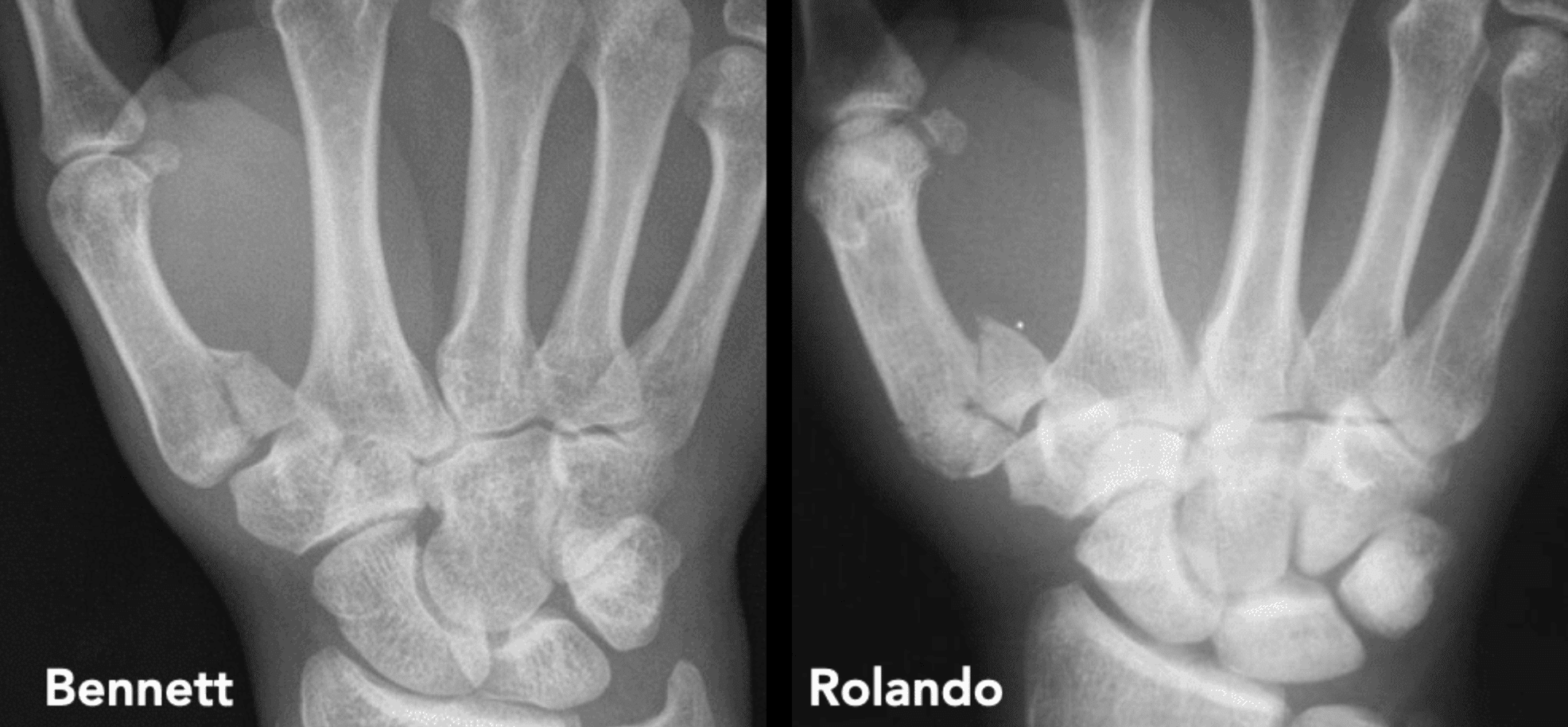

Left: Bennett fracture, a two-part intra-articular fracture-dislocation.
Right: Rolando fracture, a comminuted intra-articular fracture with three or more fragments, classically forming a Y- or T-shaped pattern.
History of the Rolando fracture
1881 – Edward Hallaran Bennett (1837–1907) described an oblique intra-articular fracture-dislocation of the base of the first metacarpal from nine pathological specimens. He presented his findings in Dublin in 1881 and published Fractures of the metacarpal bones in 1882. Bennett’s injury consisted of a two-part fracture, with a volar-ulnar articular fragment and displacement of the remaining metacarpal.
1897 – Bennett expanded on his initial presentation to the Royal Academy of Medicine in Ireland the injury introducing pathological specimens, casts, photographs and the newly available X-rays. Sir William Stokes (1839–1900) proposed that the injury should thereafter be associated with Bennett’s name.
1908 – Samuel Robinson reviewed 92 radiographically documented fractures of the first metacarpal, including 28 Bennett-type injuries. He challenged the assumption that the Bennett fracture pattern was the commonest first-metacarpal fracture.
1910 – Silvio Rolando (1873–1949) described first-metacarpal fractures in Fracture de la base du premier metacarpien et principalement sur une variété non encore décrite. Rolando reported 12 fractures of the first metacarpal encountered among port workers. Two involved the shaft and ten the base. Of the basal fractures, two were extra-articular, five were Bennett fractures, and three represented a previously undescribed intra-articular Y-shaped pattern.
In these three injuries the metacarpal base was divided into the metacarpal shaft (diaphyseal fragment); a dorsal articular fragment; and a palmar articular fragment.
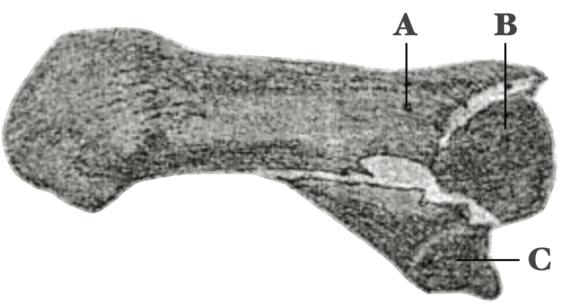
Rolando proposed that greater or more prolonged axial force than that producing a Bennett fracture could fracture the stronger dorsal portion of the metacarpal base. This created a the three-part configuration.
Original
English
Toutes les fois donc que la violence exercée au long de l’axe longitudinal du métacarpe agit plus intensément ou plus longuement, elle ourrait entraîner aussi la fracture du procès articulaire plus résistant, c’est-à-dire du procès dorsal. Dans ce cas-là il se produit une fracture en Y par laquelle l’extrémité supérieure du premier métacarpien reste partagé en trois fragments, dont deux, correspondant à la base, sont respectivement dorsal et palmaire, et l’autre correspond au corps de l’os.
Dans les trois cas de fractures en Y, le facteur étiologique est représenté dans un cas par un coup de poing fortement vibré contre la tête de l’adversaire, le pouce étant replié et serré dans la paume de la main; dans les deux autres cas par chute sur le côté radial de la main avec le pouce en adduction.
Rolando 1910; 33: 303–304
When trauma occurs to the long axis of the metacarpal, it can cause a fracture of the dorsal process. In this case, a Y shaped fracture is produced by which the proximal first metacarpal is divided into three fragments of which two parallel the base, dorsal and palmar respectively, and the other corresponds to the body of the bone.
In the 3 cases of Y fractures, the aetiologic factor is represented in one case by a blow with a closed fist, very strongly against the head of the adversary with the thumb folded and held into the palm of the hand, in the other two cases by a fall on the radial side with the thumb in adduction.
Rolando 1910; 33: 303–304
Rolando stated that the injury could not reliably be distinguished from a Bennett fracture without radiographic examination. He recognised the tendency of the fracture to redisplace and its poor functional prognosis, especially when displaced fractures were left untreated or inadequately reduced.
Original
English
Le pronostic est défavorable au point de vue fonctionnel toutes les fois que de telles fractures, (principalement quand les fragments en sont mobiles et le déplacement remarquable) sont abandonnées à elles-mêmes, ou n’ont pas reçu un traitement convenable.
The prognosis is unfavorable from the functional point of view whenever this fracture (principally when the fragments are mobile and there is noticeable displacement) is left to itself or has not received the appropriate treatment
Rolando’s original lesion was therefore a three-part Y-shaped complete intra-articular fracture, rather than simply any comminuted fracture of the first metacarpal base. He treated the injury by reduction with the thumb abducted and extended, followed by traction and plaster immobilisation.
Original
English
Il résulte de mes observations qu’il existe un type de fracture de la base du premier métacarpien, qui n’avait pas encore été décrit à ce que je sache. Cette fracture, que j’ai observé trois fois sur dix cas de fracture de la base, est consécutive à une violence qui agit suivant l’axe longitudinal du métacarpe; elle possède une forme en Y, ne peut pas se distinguer de la fracture de Bennett sans l’intervention radioscopique, et, comme celle-là, elle exige un traitement spécial.
Rolando 1910; 33: 304
I have found that there exists a type of fracture of the base of the first metacarpal that has not yet been described as far as I know. This fracture that I have noticed in 3 cases of 10 of base fractures follows an injury acting along the longitudinal axis of the metacarpal. It has a Y form and cannot be distinguished from a Bennett’s fracture without radiographic studies, and like the Bennetts’s fracture, it has to have a special kind of treatment.
Rolando 1910; 33: 304
1979 – Gelberman, Vance and Zakaib extended oblique traction techniques previously used for Bennett fractures to comminuted intra-articular fractures of the first metacarpal. The technique aimed to counter shortening and varus angulation to maintain satisfactory reduction in their small mixed series.
1991 – Ueli Büchler, Stephen M. McCollam and Claude Oppikofer reported combined treatment of severely comminuted thumb CMC fractures using intermetacarpal external fixation, limited internal fixation and bone grafting. They reported 13 displaced comminuted fractures, each with at least three articular fragments. All fractures united without secondary displacement, with grip strength averaging 81% and key pinch 88% of the contralateral hand.
1992 – Ignacio R. Proubasta reported five Rolando fractures treated by closed reduction and external fixation between the trapezium and first metacarpal. At three months all five patients were pain-free with a full range of thumb movement with no reflex sympathetic dystrophy or superficial radial nerve injury reported. Proubasta proposed external fixation as a means of maintaining reduction in severely comminuted injuries in which internal fixation was difficult.
1996 – Roy A. Meals published his English translation of Rolando’s original 1910 paper helping reintroduce the original description and its specific three-part Y-shaped morphology to the English-language literature
2014 – Modern reviews by Sood and Granick, and Mahoney and colleagues, emphasized that the eponymous term had broadened to include multi-fragmentary intra-articular fractures of the first-metacarpal base beyond Rolando’s original three-part injury description.
Associated Persons
- Edward Hallaran Bennett (1837–1907)
- Silvio Rolando (1873-1949)
Alternative names
- Rolando’s fracture
- Intra-articular thumb metacarpal base fracture with three or more segments
References
Original articles
- Rolando S. Fracture de la base du premier metacarpien et principalement sur une variété non encore décrite. La Presse Médicale 1910; 33: 303–304. [Translated Meals RA. Fracture of the base of the first metacarpal and a variation that has not yet been described. Clin Orthop Relat Res. 1996 Jun;(327):4-8]
Review articles
- Gedda KO. Studies on Bennett’s fracture; anatomy, roentgenology, and therapy. Acta Chir Scand Suppl. 1954;193:1-114.
- Gelberman RH, Vance RM, Zakaib GS. Fractures at the base of the thumb: treatment with oblique traction. J Bone Joint Surg Am. 1979 Mar;61(2):260-2.
- Howard FM. Fractures of the basal joint of the thumb. Clin Orthop Relat Res. 1987 Jul;(220):46-51.
- Buchler U, McCollam SM, Oppikofer C. Comminuted fractures of the basilar joint of the thumb: combined treatment by external fixation, limited internal fixation, and bone grafting. J Hand Surg Am. 1991 May;16(3):556-60.
- Proubasta IR. Rolando’s fracture of the first metacarpal. Treatment by external fixation. J Bone Joint Surg Br. 1992 May;74(3):416-7
- Soyer AD. Fractures of the base of the first metacarpal: current treatment options. J Am Acad Orthop Surg. 1999 Nov-Dec;7(6):403-12.
- Edmunds JO. Traumatic dislocations and instability of the trapeziometacarpal joint of the thumb. Hand Clin. 2006 Aug;22(3):365-92
- Sood A, Granick MS. Rolando fracture. Eplasty. 2014 Jun 6;14:ic16
- Mahoney M, Marsland D, Garagnani L, Sauvé P. Rolando and his fracture. Trauma. 2015;17(1):24-28.
- Feletti F, Varacallo M. Rolando Fractures. 2021 Jul 18. In: StatPearls [Internet]. Treasure Island (FL): StatPearls Publishing; 2022
- Vajuhudeen Z. Rolando Fracture. Radiopaedia
- Cadogan M. Rolando fracture. Eponym A Day. Instagram
eponymictionary
the names behind the name

MBChB University of Manchester, PG Dip (SEM) University of Bath. Keen interest in Sport and Exercise Medicine. Currently working in emergency medicine in Perth, WA and intend to train as an ACSEP Registrar.

BA MA (Oxon) MBChB (Edin) FACEM FFSEM. Emergency physician, Sir Charles Gairdner Hospital. Passion for rugby, medical history, medical education, and asynchronous learning #FOAMed evangelist. Editor-in-Chief and Webmaster of Life in the Fast lane, LITFL | On Call 4e| Eponyms | Books | Horology |