


Bennett fracture
Bennett fracture is an unstable two-part intra-articular fracture-dislocation of the base of the first metacarpal involving the thumb carpometacarpal (CMC) joint. A volar-ulnar articular fragment remains relatively aligned with the trapezium while the remainder of the metacarpal subluxates proximally and dorsoradially under deforming muscular forces.
The classic mechanism is an axial load transmitted through a partially flexed thumb, such as a fall onto the thumb or impact through a clenched fist.
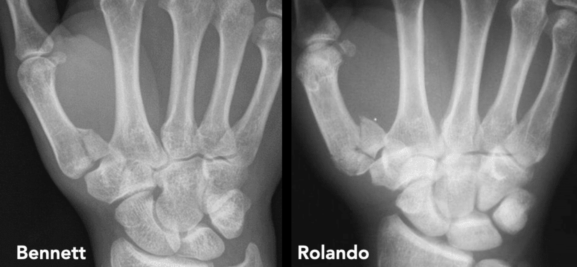

Left: Bennett fracture, a two-part intra-articular fracture-dislocation with a volar-ulnar articular fragment remaining associated with the trapezium and displacement of the metacarpal shaft.
Right: Rolando fracture, a comminuted intra-articular fracture with three or more fragments, classically forming a Y- or T-shaped pattern.
Definition
Bennett fracture: partial intra-articular fracture-dislocation of the base of the first metacarpal. The volar-ulnar marginal fragment remains associated with the trapezium while the remainder of the metacarpal is displaced at the first CMC joint.
Mechanism and anatomy
The usual mechanism is axial compression through a flexed first metacarpal. The force produces an oblique intra-articular fracture through the volar-ulnar base. The residual metacarpal shaft and articular component tend to displace proximally, dorsally and radially.
The volar-ulnar fracture fragment remains relatively fixed to the trapezium by its ligamentous attachments, while the metacarpal shaft subluxates dorsoradially and proximally. The anterior oblique ligament has traditionally been considered the principal attachment, although modern studies suggest a possibly dominant role for the CMC ulnar collateral ligament.
Clinical features
Patients typically present following trauma with:
- pain and tenderness centred over the thumb CMC joint;
- swelling and ecchymosis at the base of the thumb;
- painful restriction of thumb movement;
- reduced pinch and grip strength; and
- occasionally an obvious deformity when displacement is substantial.
The skin and neurovascular status should be documented, and associated fractures or ligamentous injuries considered in higher-energy trauma.
Imaging
Dedicated thumb radiographs usually establish the diagnosis. Recommended views include a true AP view of the thumb (Robert view), lateral and oblique views. A 30° pronated view may better demonstrate the first CMC articulation. CT is useful when the fracture configuration is uncertain, when comminution is suspected or for operative planning.
Management
The objectives are to restore CMC joint congruity, reduce the metacarpal subluxation and maintain a stable reduction while the fracture heals.
Initial management
Immobilise in a thumb-spica splint, provide analgesia and elevation, document neurovascular status and arrange appropriate hand/orthopaedic follow-up. The thumb should not be repeatedly manipulated without a definitive reduction plan.
Non-operative treatment
Closed reduction and thumb-spica immobilisation may be considered for an essentially nondisplaced fracture that is stable after reduction and can be monitored closely. In practice, many displaced Bennett fractures are unstable because the muscular deforming forces tend to reproduce the CMC subluxation.
Operative treatment
For displaced or unstable injuries, options include:
- closed reduction with percutaneous K-wire fixation;
- open reduction and internal fixation with screws or wires; and
- less commonly other fixation strategies according to fragment size and fracture morphology.
A residual articular step of around 1 mm is frequently used as a practical threshold when assessing reduction, although the evidence does not establish a single absolute cutoff applicable to every patient.
| Bennett fracture | Rolando fracture | |
|---|---|---|
| Pattern | Two-part intra-articular fracture-dislocation | Comminuted intra-articular fracture |
| Articular fragments | Single volar-ulnar marginal fragment plus metacarpal shaft | Three or more fragments |
| Classic shape | Oblique fracture through volar-ulnar base | Y- or T-shaped |
| CMC stability | Unstable | Usually highly unstable |
| Mechanism | Axial load through flexed thumb | Similar axial load Usually greater energy/comminution |
| Fixation | Often amenable to percutaneous pinning or screw fixation | Fixation dictated by fragment size. Reconstruction may be more difficult |
| Prognosis | Generally favourable if reduction maintained | Less predictable due to comminution |
NOTE: Bennett = two-part intra-articular fracture-dislocation; Rolando fracture = comminuted three-or-more-part intra-articular fracture.
History of the Bennett fracture
1879 – Later accounts state that Bennett first described the fracture at the British Medical Association meeting in Cork in 1880. However Robert O’Rahilly’s 1948 review found no evidence for this. The Cork meeting actually occurred in 1879, and Bennett’s presentations concerned femoral fractures, Colles fracture and compound fractures but nothing relating to the first metacarpal.
November 12, 1881 – Edward Hallaran Bennett (1837–1907) presented a series of healed metacarpal fractures to the Pathological Society of Dublin. Of nine metacarpal specimens, five involved the first metacarpal and showed a recurring oblique fracture through its base. All had been obtained from the dissecting room, fourteen years before Röntgen’s discovery of X-rays.
Bennett considered the pattern previously undescribed and recognised that the major metacarpal component underwent dorsal subluxation. He also noted that the injury could heal with little obvious deformity yet leave the thumb painful and functionally impaired for months.
1882 – Published Fractures of the metacarpal bones in the Dublin Journal of Medical Science. Within his larger series of metacarpal injuries, Bennett identified a recurring oblique intra-articular fracture at the base of the thumb metacarpal which clinically resembled a dislocation.
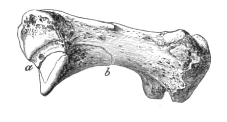
The fracture passes obliquely (a, b in woodcut) through the base of the bone, detaching the greater part of the articular facet with that piece of the bone supporting it, which projects into the palm.
Bennett 1882
1886 – Bennett published specifically on the lesion in On Fracture of the Metacarpal Bone of the Thumb‘, reinforcing its status as a distinct injury pattern.
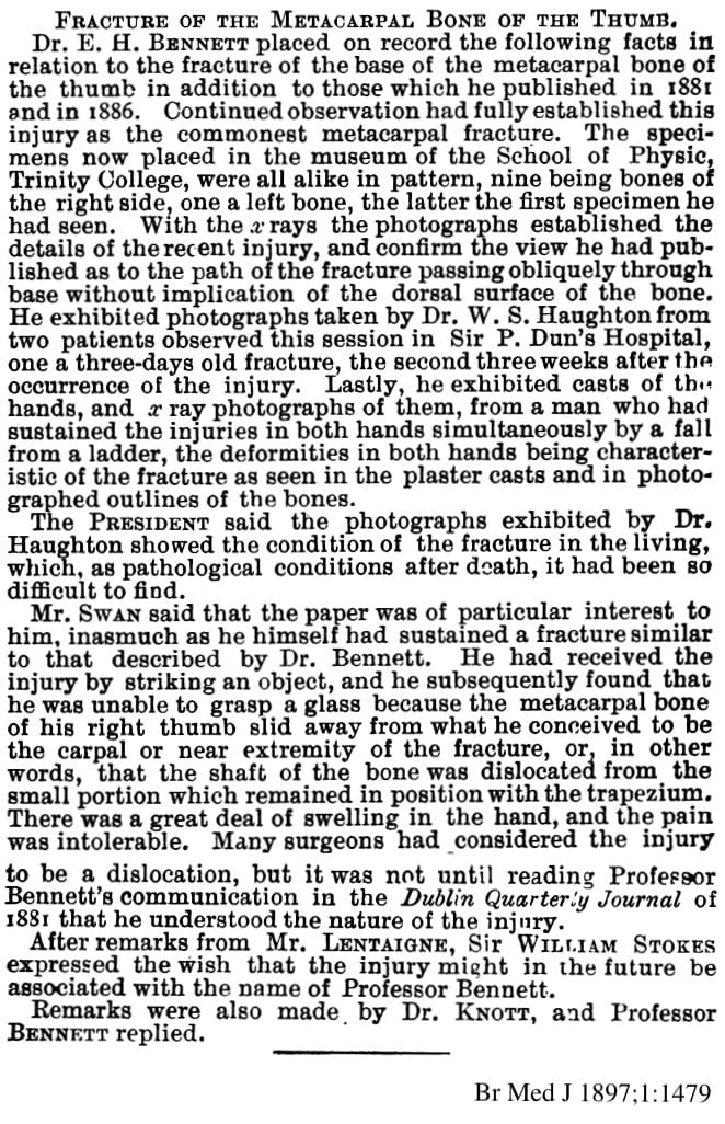
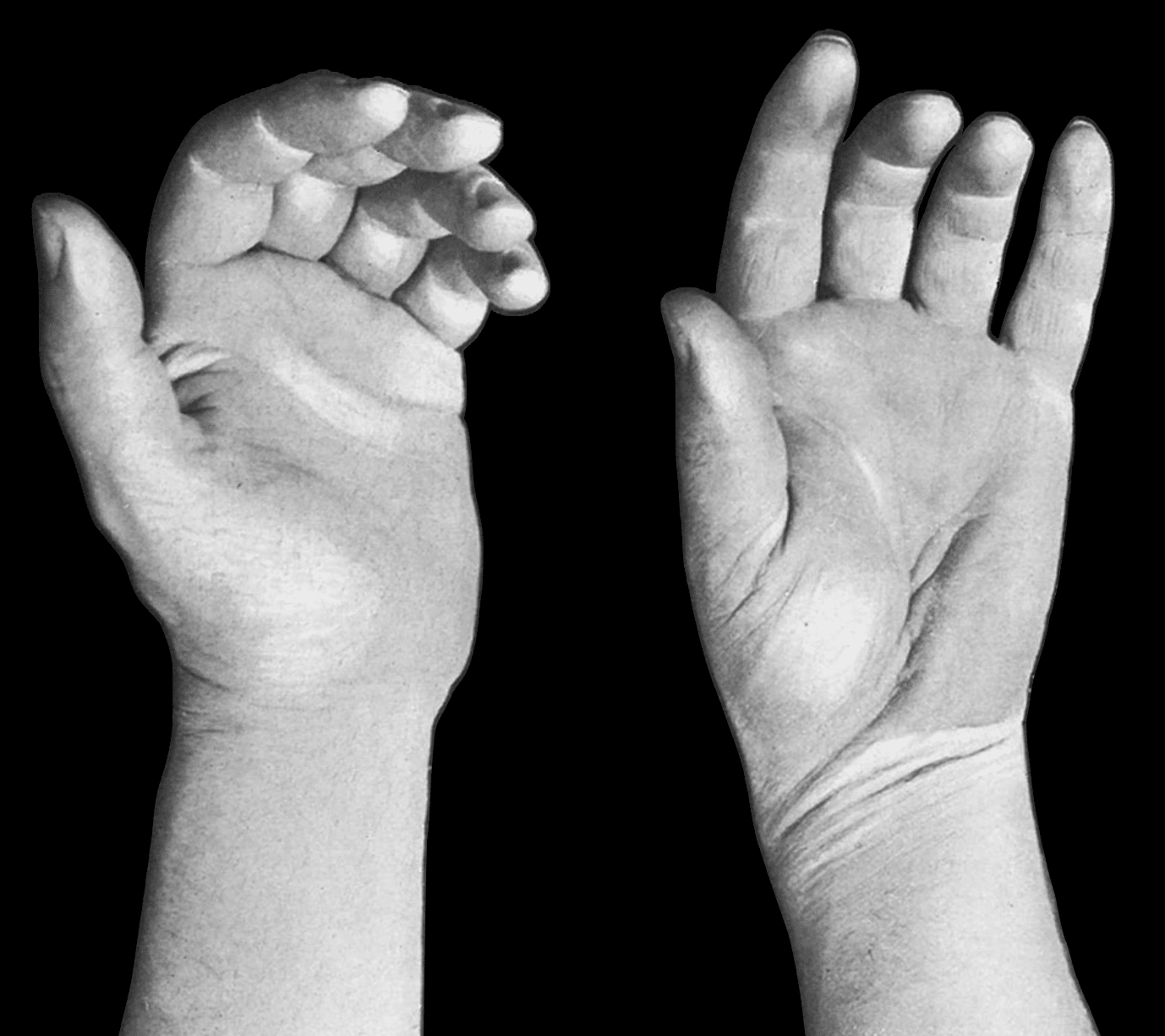
1897 – Bennett presented to the surgical section of the Royal Academy of Medicine in Ireland. He combined his pathological specimens with casts, photographs and X-rays of living patients taken by Dr W. S. Haughton. The radiographs demonstrated the fracture in vivo and confirmed the oblique fracture path Bennett had inferred from anatomical specimens.
Following the presentation and discussion:
Sir William Stokes expressed the wish that the injury might in the future be associated with the name of Professor Bennett.
Br Med J 1897;1:1479
Transcript of 1897 Presentation to the Royal Academy of Medicine in Ireland
1908 – Bennett’s original conclusion was based on pathological specimens and was the dominant fracture in the small collection available to him. Bennet suggested that it was be the commonest fracture of the first metacarpal.
Samuel Robinson challenged this in The Bennett fracture of the first metacarpal bone; diagnosis and treatment. Robinson reviewed 92 radiographically documented fractures of the first metacarpal and found that only 28 (30%) were Bennett-type intra-articular fractures. He concluded that radiography was needed to establish whether the CMC joint was involved and whether subluxation was present.
Robinson also reviewed earlier surgical authors, including Malgaigne, Hulke and Hamilton, who had discussed metacarpal fractures and their relative frequency. He identified no earlier specific description of the characteristic intra-articular fracture-subluxation described by Bennett.
1910 – Silvio Rolando (1873–1949) described 12 first-metacarpal fractures in Fracture de la base du premier metacarpien et principalement sur une variété non encore décrite. This included three injuries in which the base was divided into dorsal and palmar articular fragments plus the metacarpal shaft, producing a Y-shaped fracture. Rolando concluded that this pattern could not reliably be distinguished from Bennett fracture without radiography.
1936 – French radiologist Paul Robert described dedicated radiographic positioning of the trapeziometacarpal joint. Robert demonstrated forced supination and forced pronation positions and favoured the pronated technique as the more practical and reproducible method. This projection subsequently became known as the Robert view and remains useful for assessment of the first CMC joint and fractures of the first metacarpal base.
1952 – Radiologist Lars Billing and surgeon Karl-Ove Gedda (1909–2003) acknowledged the Robert view as useful for demonstrating radial displacement. However, in their paper on Roentgen examination of Bennett’s fracture they demonstrated that a true lateral projection was necessary for accurate assessment of the displacement and reduction of a Bennett fracture.
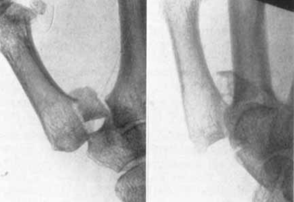
1952 – K.-O. Gedda and Erik Moberg described open reduction and osteosynthesis of Bennett fractures through a volar approach at the Sahlgrenska hand surgery service. Their work reflected a shift toward direct restoration of the articular surface and stable internal fixation rather than reliance on plaster immobilisation alone.
1954 – Gedda published the 114-page monograph Studies on Bennett’s fracture; anatomy, roentgenology, and therapy, synthesising anatomical investigation, standardised X-ray assessment and operative treatment.
1990 – Kjær-Petersen, Langhoff and Andersen reviewed 41 Bennett fractures with a median follow-up of 7.3 years. Patients whose fractures healed with <1 mm residual displacement were more frequently asymptomatic and had substantially less radiographic CMC osteoarthritis than those with residual displacement. Despite this, late symptoms were generally mild. The authors advocated accurate reduction and stable fixation, using open reduction when satisfactory alignment could not otherwise be maintained.
1994 – Timmenga et al reviewed 18 first-metacarpal base fractures after a mean 10.7 years. Better reduction correlated with less severe radiographic osteoarthritis, although degenerative change developed in almost all patients, including most with apparently exact reduction, and did not correlate with symptoms. Treatment method itself was not associated with arthritis or joint deformity. Interpretation is limited by the small, heterogeneous cohort, which included comminuted and Rolando fractures as well as classic Bennett injuries.
2003 – Lutz, Sailer and colleagues compared closed reduction with transarticular K-wire fixation against open reduction with lag-screw fixation in 32 Gedda type I Bennett fractures followed for a mean of seven years. When articular reduction to ≤1 mm could be achieved, the method of fixation did not significantly influence clinical outcome or radiographic osteoarthritis. Percutaneous fixation produced more adduction deformity, particularly when wire placement contributed to loss of reduction, but without inferior functional results. The authors favoured closed reduction and pinning for reducible large-fragment injuries and reserved open fixation for irreducible fractures.
2019 – Kang, Behn, Messana and Ladd developed a cadaveric biomechanical model that reproducibly generated displaced intra-articular palmar beak fractures. Contrary to the traditional model, the anterior oblique ligament was thin and only partly attached to the beak fragment, whereas the more robust ulnar collateral ligament remained attached in its entirety. Radial abduction and pronation reduced the experimental fracture, while extension and tensioning of the anterior oblique ligament increased displacement. The findings challenged the traditional explanation of Bennett fracture ligamentous anatomy, although they derived from a small cadaveric model.
Eponymic foundations
The Bennett fracture is an unusual exception to Stigler’s law of eponymy where an eponym has been correctly attributed to the physician who first characterised the lesion.
Edward Hallaran Bennett recognised the characteristic fracture-dislocation from pathological specimens and first presented it to the Pathological Society of Dublin in 1881. Bennett did not name the injury after himself; in 1897 Sir William Stokes explicitly proposed that the lesion should thereafter be associated with Bennett’s name. No convincing earlier description of the characteristic intra-articular fracture-dislocation has been identified.
Bibliographic note. Paul Robert and the “M. Robert” error
The dedicated trapeziometacarpal projection later known as the Robert view was described in 1936 by French radiologist Paul Robert. Later bibliographies cite the author as “M. Robert” and bibliographic databases have consequently interpreted M. as a first initial. In the original French context, however, M. = Monsieur. The author was Paul Robert, and the citation should therefore be rendered Robert P., not “Robert M.”
Associated Persons
- Edward Hallaran Bennett (1837-1907)
- Sir William Stokes (1839–1900)
- Silvio Rolando (1873-1949)
Alternative names
- Bennett’s fracture
- Bennett fracture-dislocation
- Bennett fracture-luxation
- Two-part intra-articular fracture of the first metacarpal base
References
Original articles
- Bennett EH. Fractures of the metacarpal bones. Dublin J Med Sci 1882; 73: 72-75
- Bennett EH. On fracture of the metacarpal bone of the thumb. Br Med J 1886; 2: 12-13
- Bennett EH. Report of a Meeting of the Royal Academy of Medicine in Ireland 14 May 1897. Br Med J 1897; 1: 1479
- Robinson S. The Bennett fracture of the first metacarpal bone; diagnosis and treatment. Boston Med Surg J. 1908;158:275–280.
- Rolando S. Fracture de la base du premier metacarpien et principalement sur une variété non encore décrite. La Presse Médicale 1910; 33: 303–304.
- Robert P. La radiographie de l’articulation trapézo-métacarpienne. Les arthroses de cette jointure. Société de Radiologie Médicale de France, Bulletins. 1936;24:687–689. [Robert P. The classic: Radiography of the trapeziometacarpal joint. Degenerative changes of this joint. 1936. Clin Orthop Relat Res. 2014 Apr;472(4):1095-6.]
- Billing L, Gedda KO. Roentgen examination of Bennett’s fracture. Acta Radiol (Stockh). 1952 Dec;38(6):471-6.
- Gedda KO, Moberg E. Open reduction and osteosynthesis of the so-called Bennett’s fracture in the carpo-metacarpal joint of the thumb. Acta Orthop Scand. 1952;22(1-4):249-57.
- Gedda KO. Studies on Bennett’s fracture; anatomy, roentgenology, and therapy. Acta Chir Scand Suppl. 1954;193:1-114.
- Kjaer-Petersen K, Langhoff O, Andersen K. Bennett’s fracture. J Hand Surg Br. 1990 Feb;15(1):58-61.
- Timmenga EJ, Blokhuis TJ, Maas M, Raaijmakers EL. Long-term evaluation of Bennett’s fracture. A comparison between open and closed reduction. J Hand Surg Br. 1994 Jun;19(3):373-7.
- Lutz M, Sailer R, Zimmermann R, Gabl M, Ulmer H, Pechlaner S. Closed reduction transarticular Kirschner wire fixation versus open reduction internal fixation in the treatment of Bennett’s fracture dislocation. J Hand Surg Br. 2003 Apr;28(2):142-7.
- Ladd AL. Guest editorial: The Robert’s view: a historical and clinical perspective. Clin Orthop Relat Res. 2014 Apr;472(4):1097-100
- Kang JR, Behn AW, Messana J, Ladd AL. Bennett Fractures: A Biomechanical Model and Relevant Ligamentous Anatomy. J Hand Surg Am. 2019 Feb;44(2):154.e1-154.e5.
Review articles
- Nickson C. Gordon Bennett! Bone and Joint Bamboozler 004
- Baba Y. Bennett fracture. Radiopaedia
- Brownlie C, Anderson D. Bennett fracture dislocation – review and management. Aust Fam Physician. 2011 Jun;40(6):394-6.
- Goru P, Haque S, Verma GG, Mustafa A, Ebinesan A. Bennett’s Fracture Management: A Systematic Review of Literature. Cureus. 2022 Nov 10;14(11):e31340.
eponymictionary
the names behind the name

MBChB University of Manchester, PG Dip (SEM) University of Bath. Keen interest in Sport and Exercise Medicine. Currently working in emergency medicine in Perth, WA and intend to train as an ACSEP Registrar.

BA MA (Oxon) MBChB (Edin) FACEM FFSEM. Emergency physician, Sir Charles Gairdner Hospital. Passion for rugby, medical history, medical education, and asynchronous learning #FOAMed evangelist. Editor-in-Chief and Webmaster of Life in the Fast lane, LITFL | On Call 4e| Eponyms | Books | Horology |