![]()
Roth spots
Description
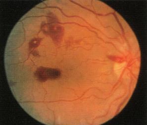
Roth spots: Retinal haemorrhages with white or pale centres, commonly associated with subacute bacterial endocarditis and immune complex mediated vasculitis.
These spots may also be found in cases of leukaemia, diabetes, hypertensive and HIV retinopathy
History
1872 – Moritz Roth (1839-1914) originally described retinal white spots and separate scattered haemorrhages (retinal red spots) in close proximity to the optic disc in patients with septicaemia (retinitis septica). Roth originally attributed the retinal spots to disseminated foci of bacterial abscesses; however, the white-centre of retinal haemorrhages in Roth spots are currently thought to be fibrin-platelet plugs.
- Roth M. Über Netzhautaffectionen bei Wundfiebern. I. Die embolische Panophthalmitis. Deutsche Zeitschrift für Chirurgie, 1872; 1(5): 471-484
1876 – Moritz Litten (1845-1907) first used the term ‘Roth spot’ article (published 1878) to describe the current understanding of Roth spots, when he observed retinal haemorrhages with a white centre in patients with endocarditis. Sometime referred to as Litten spots/sign.
- Litten M. Uber akute maligne endokarditis und die dabei vorkommenden retinalveränderungen. Charité-Annalen 1876; III: 137-172. (published 1878)
Associated Persons
- Moritz Roth (1839-1914)
- Moritz Litten (1845-1907)
Alternative names
- Litten sign, Litten spots
- Roth’s spots
References
- Roth M. Über Netzhautaffectionen bei Wundfiebern. I. Die embolische Panophthalmitis. Deutsche Zeitschrift für Chirurgie, 1872; 1(5): 471-484
- Litten M. Uber akute maligne endokarditis und die dabei vorkommenden retinalveränderungen. Charité-Annalen 1876; III: 137-172. (published 1878)
- Khawly JA, Pollock SC. Litten’s Sign (Roth’s Spots) in Bacterial Endocarditis. Arch Ophthalmol. 1994;112(5):683-684

eponymictionary
the names behind the name
Doctor in Australia. Keen interest in internal medicine, medical education, and medical history.