Terry’s Nails
Description
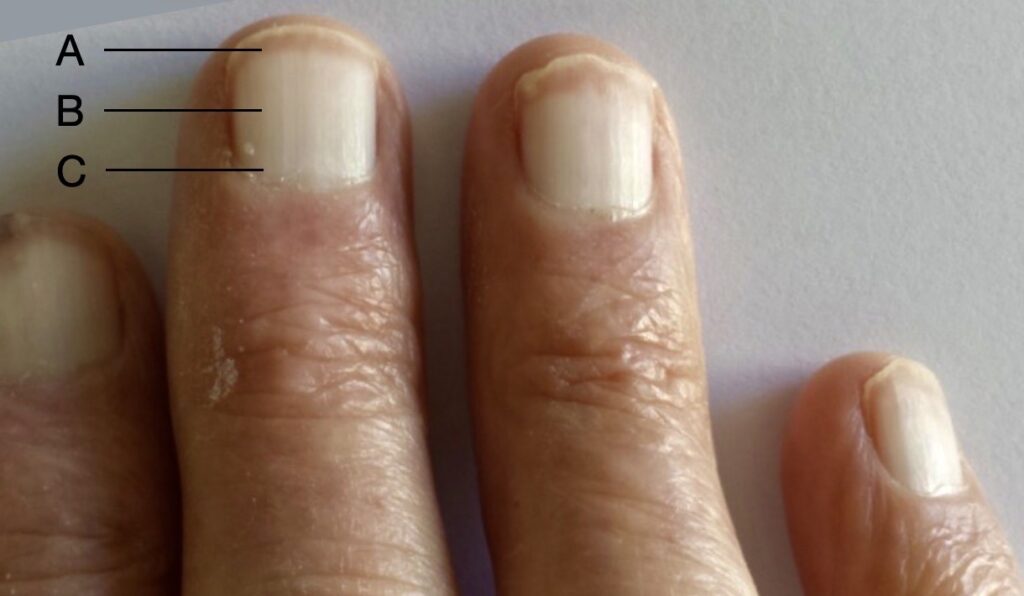
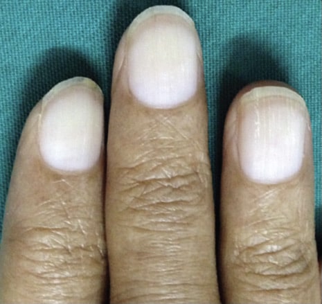
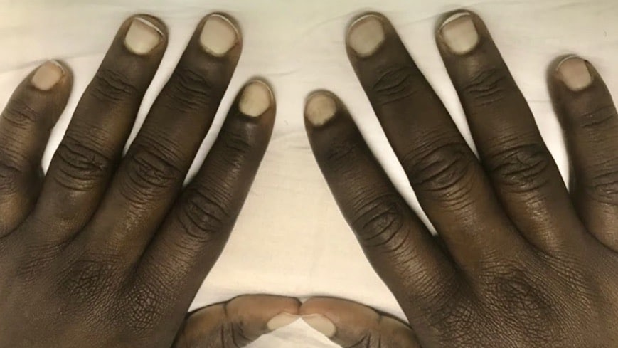
Terry’s nails are a type of apparent leukonychia, characterised by ground glass opacification of almost the entire nail; distal narrow band of normal, pink nail bed; and often with obliteration of the lunula.
The narrow pink/ brown segment (0.5 – 3mm) of the distal border before the lunula indicates normal nail bed tissue. Opacity varies in degree and distribution: the most severe change is a proximal white nail with a dark band distally as shown below. The condition is usually bilaterally symmetrical, with a tendency to be more marked in the thumb and forefinger.
This sign is also found in systemic diseases such as chronic congestive heart failure (p <0.01), adult onset diabetes mellitus (p< 0.001) but also pulmonary tuberculosis, rheumatoid arthritis, convalescent viral hepatitis, disseminated sclerosis, renal failure and metastatic cancer. Terry’s nail is part of normal ageing and the above systemic diseases “age” the nails quicker than normal.
Terry nails should alert the clinician to the possibility of an underlying systemic disease, especially advanced liver disease

A: Distal thin pink-brown transverse band, 0.5-3mm wide, not obscured by venous congestion
B: white or light pink nail
C: lunula may or may not be present
Whitening of the fingernail, often reported anecdotally, was not associated with a systemic disorder until 1954, when Richard Barratt Terry (1914-1960) reported finding a white nail bed, showing ground-glass opacity, not affected by venous congestion, and indistinguishable from the lunula, with a distal band of normal pink, in 82 of 100 cirrhotic patients.
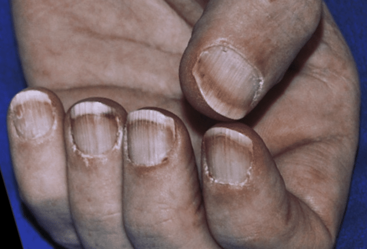
Differential diagnosis for Terry’s nails includes half-and-half nails (Lindsay nails with only only about half of the proximal nail bed is opacified), Muehrcke lines (paired, white, transverse lines that typically spare the thumbnail), and true leukonychia totalis/partialis
History of Terry’s nails
1954 – Initially described by Richard Terry in patients with hepatic cirrhosis (sign demonstrated in 82 of 100 cirrhotic patients). Terry investigated “Opacity of the nail bed causing apparent whiteness of the finger-nails, and its occurrence in cirrhosis of the liver and some other conditions.” The whitened appearance of the nail due to underlining defects of the nail bed was termed ‘apparent leuconychia’.
Fully developed white nails exhibit a ground-glass-like opacity of almost the entire nail bed. It extends from the base of the nail, where the lunula is indistinguishable, to within one or two millimetres of the distal border of the nail bed, leaving a distal zone of normal pink. The condition is bilaterally symmetrical, with a tendency to be more marked in the thumb and forefinger.
Terry 1954
The pathophysiology remains underdetermined but currently thought to be due to changes in nail bed vascularity secondary to overgrowth of connective tissue. Nail bed tissue biopsy confirm microvascular involvement showing telangiectasias in the upper dermis of the distal band.
These nails are common in cirrhosis of the liver and may fairly be added to the list of non-specific physical signs thereof. Their diagnostic value in cirrhosis is limited, since they occur in other conditions, but they are occasionally most helpful in suggesting or corroborating the diagnosis.
Terry 1954

1984 – Mark Holzberg and H. Kenneth Walker examined the fingernails of 512 consecutive hospital inpatients were examined. Based on their findings, they redefined Terry’s original criteria:
- Distal thin pink-to-brown transverse band, 0.5-3.0mm in width (rather than 1-2mm)
- Decreased venous return not obscuring the distal band
- White or light pink proximal nail (rather than white ground-glass appearance)
- Lunula possibly absent (rather than always absent)
- At least 4 of 10 nails with the above criteria
Terry’s nails (with modified criteria) were found in 25·2%.
The nail abnormality was associated with cirrhosis; chronic congestive heart failure; adult-onset diabetes mellitus; and age. In younger patients the nail disorder was associated with an increased risk of systemic disease. Tissue biopsy showed that the nail abnormality was due to distal telangiectasias.
1992 – Park et al used the updated diagnostic criteria, and reviewed the fingernails in 444 medical inpatients with chronic systemic disease. They found 30.6% had Terry nails. There were statistically significant associations with cirrhosis (57%); congestive heart failure (51.5%); diabetes mellitus (49%); and non statistically significant associations with chronic renal failure (19%); and cancer (18%)
Increased incidence with age. The average number of nails affected per patient tended to be higher in frequency close to the thumb and 28.7% patients had all nails affected

Associated Persons
- Richard Barratt Terry (1914-1960)
Alternative names
- Terry nails
Other eponymous nail signs
References
Original articles
- Burrows MT. The significance of the lunula of the nail. The Anatomical Record 1917; 12(1): 161-166
- Howard et al. Leukonychia in three generations. Journal of cutaneous diseases 1917; 35: 559
- Pernet G. Case of Multiple Leuconychia Striata, associated with Leuconychia Totalis of One Thumb Nail. Proc R Soc Med. 1919; 12(Dermatol Sect): 28‐29.
- Weis. Leukonychia. Journal of cutaneous diseases 1918; 36: 594-95.
- Kleeberg J. Flat finger-nails in cirrhosis of the liver. Lancet. 1951;2(6676):248‐249.
- Terry R. White nails in hepatic cirrhosis. Lancet. 1954; 266(6815): 757‐759.
Review articles
- Morey DA, Burke JO. Distinctive nail changes in advanced hepatic cirrhosis. Gastroenterology 1955; 29: 258-61.
- Stewart WK, Raffle EJ. Brown nail-bed arcs and chronic renal disease. Br Med J. 1972; 1(5803): 784‐786.
- Holzberg M, Walker HK. Terry’s nails: revised definition and new correlations. Lancet. 1984; 1(8382): 896‐899
- Park KY, Kim SW, Cho JS. Research on the frequency of Terry’s nail in the medical inpatients with chronic illnesses. Korean J Dermatol 1992; 30: 864–870
- Nia AM, Ederer S, Dahlem KM, Gassanov N, Er F. Terry’s nails: a window to systemic diseases. Am J Med. 2011; 124(7): 602‐604.
- Lakshmi B S, Ram R, Kumar V S. Terry’s nails. Indian J Nephrol 2015; 25: 184
- Pitukweerakul S, Pilla S. Terry’s Nails and Lindsay’s Nails: Two Nail Abnormalities in Chronic Systemic Diseases. J Gen Intern Med. 2016;31(8):970.
- Witkowska AB, Jasterzbski TJ, Schwartz RA. Terry’s Nails: A Sign of Systemic Disease. Indian J Dermatol. 2017; 62(3): 309‐311.
- Flores L, Moreira H, Pinto MJ, Andrade C, Friões F. Terry’s nails, tracking an underneath disease. Postgrad Med J. 2019; 95(1125): 405.
- Meegada S, Verma R. Terry’s nails. Clin Case Rep. 2020; 8(2): 404‐405.
- Cadogan M. Terry’s nails, Eponym A Day. instagram
[cite]

eponymictionary
the names behind the name
Graduated from Southampton Medical School in 2017 with BMBS. Working in Sir Charles Gairdner Hospital Emergency Department in Perth, Australia.


I have a very Severe case of terry’s nails. Was diagnosed at around 30 years of age but the nails have displayed this appearance as long as I can recall back into childhood at least from age 9. They are also thick, strong and grow very quickly. Have a diagnosis of RA, Reynauds, and other undiagnosed issues but no liver, heart or major organ involvement.I would be happy to share a photo