![]()
Ultrasound Case 013
Presentation
23 year old female presents with lower and then diffuse abdominal pain with transient hypotension and bradycardia. Urine pregnancy test is positive.
View 2
View 3

View 4

Describe and interpret these scans
IMAGE INTERPRETATION
Image 1: Right upper quadrant FAST view with free fluid.
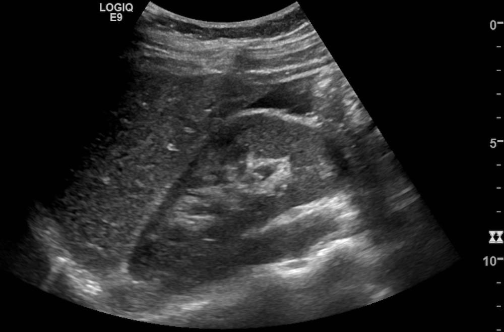
Image 2: Longitudinal pelvic view with bladder at the left of the screen, and an empty uterus in the center of the screen with free fluid and dependent clotted blood in the Pouch of Douglas.
Image 3: Longitudinal pelvic view further to the right with a heterogeneous complex cystic mass behind the uterus.
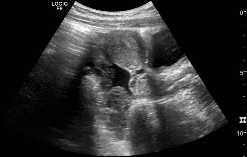
Image 4: Transverse pelvic view with an empty uterus adjacent to the anterior abdominal wall with a heterogeneous cystic mass in the right adnexa and free fluid with dependent heterogeneous clotted blood in the Pouch of Douglas.
CLINICAL CORRELATION
Sonographic evidence concerning for ruptured ectopic pregnancy, proven on laporoscopy.
[cite]
TOP 100 ULTRASOUND CASES