![]()
Ultrasound Case 018
Presentation
An 18 year old rugby player presents after after an accidental kick to the face. He has a severe periorbital haematoma and you are worried about globe injury but cannot pry his eyelids apart enough to assess the anterior chamber or pupillary function.
Can ultrasound help?
View 2
View 3
Describe and interpret these scans
IMAGE INTERPRETATION
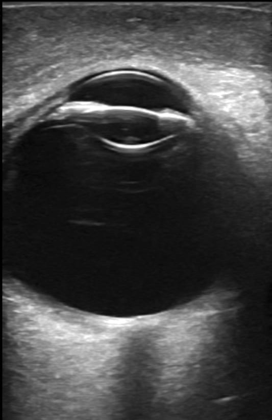
Image 1: Transverse ocular view. Scanning through the swollen, closed upper eye lid. The pre-ocular soft tissue is thickened. The cornea is seen and the anterior chamber is anechoic and intact. The iris appears normal and the lens not dislocated. There is no retinal detachment and the optic nerve is seen. No retro orbital mass is seen.
Image 2: This clip shows the same normal globe deep to the periorbital haematoma.
Image 3: Pupillary view. To get this view one needs to take an ultrasound slice through the eye in the plane of the iris. Get the patient to look downwards. Place the transducer transversely across the upper eyelid and angle it downward so the body of the transducer pushes against the upper orbital ridge and the beam is directed inferiorly toward the inferior orbital margin. The size and shape of the pupil can be assessed. Now shine a light into the opposite open eye and see if the pupil reacts consensually – as it does in this case.
CLINICAL CORRELATION
Periorbital haematoma with no injury to the globe
Assessing the globe when there is a large periorbital haematoma is difficult. Forcibly prying the eyelids open is painful, can be harmful if there is a penetrating ocular injury. It can be avoided if ultrasound is used.
We use a sterile probe cover and ultrasound gel which is placed onto the closed upper eyelid. The examination is done with the transducer horizontally across the lid. Fanning the transducer up and down allows the entire globe to be assessed.
Ocular movements can be assessed through the closed lid – hold the transducer still and get the patient to look up left and right, mid left and right, and down left and right.
Structurally the globe is assessed beginning anteriorly and working backward. Anterior orbital soft tissues, cornea, anterior chamber – content and depth, iris and lens, posterior chamber, retina, optic nerve and finally posterior periorbital tissues.
Function can be assessed by visualizing a consensual pupillary response as described above.
[cite]
TOP 100 ULTRASOUND CASES

An Emergency physician based in Perth, Western Australia. Professionally my passion lies in integrating advanced diagnostic and procedural ultrasound into clinical assessment and management of the undifferentiated patient. Sharing hard fought knowledge with innovative educational techniques to ensure knowledge translation and dissemination is my goal. Family, wild coastlines, native forests, and tinkering in the shed fills the rest of my contented time. | SonoCPD | Ultrasound library | Top 100 | @thesonocave |