Ultrasound Case 069
Presentation
A 26 year old woman describes sudden severe left iliac fossa pain late in the evening prior to presentation. On the morning of presentation she complained of bilateral shoulder tip pain and had a presyncopal episode on standing up. She is not pregnant.
View 2: Fanning through Morrison’s pouch
View 3: Fanning through the left upper quadrant
View 4: Longitudinal view fanning and sweeping through the pelvis
Describe and interpret these scans
IMAGE INTERPRETATION
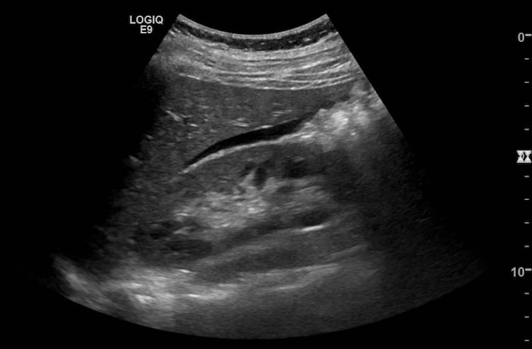
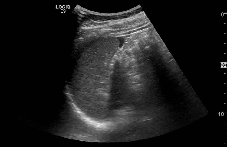
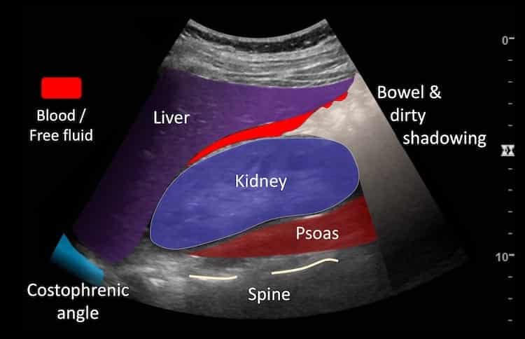
Image 1 & 2: These are images taken fanning through Morrison’s pouch, the first from more anteriorly and the second more laterally in the right upper quadrant of the abdomen.
Both show a moderate amount of anechoic fluid in Morrison’s pouch, with “sharp angles” – as is typical for free fluid fitting into the spaces between rounded organs.
Image 3: Fanning through the left upper quadrant .
The spleen is seen with free fluid at its tip and a thin strip between the diaphragm and spleen. The loop begins showing the spleen and stomach and then shows the left kidney as the transducer fans posteriorly.
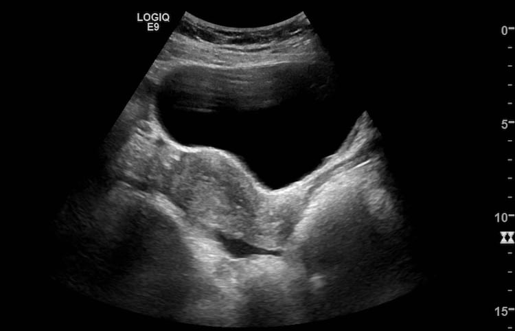
Image 4: Longitudinal view fanning and sweeping through the pelvis from right to left.
The right paracolic gutter is seen first filled with some free fluid. The bladder and uterus then come into view and fluid is seen in the Pouch of Douglas. Next the left iliac fossa is explored and a rounded heterogeneous mas is seen – clot surrounding the ovary and the ruptured corpus luteum. Finally free fluid is seen in the left paracolic gutter.
Image 5: Still image of RUQ
Image 6 Still image of LUQ
Image 7 Still image of pelvis longitudinal midline.
[cite]
TOP 100 ULTRASOUND CASES
An Emergency physician based in Perth, Western Australia. Professionally my passion lies in integrating advanced diagnostic and procedural ultrasound into clinical assessment and management of the undifferentiated patient. Sharing hard fought knowledge with innovative educational techniques to ensure knowledge translation and dissemination is my goal. Family, wild coastlines, native forests, and tinkering in the shed fills the rest of my contented time. | SonoCPD | Ultrasound library | Top 100 | @thesonocave |


