Ultrasound Case 074
Presentation
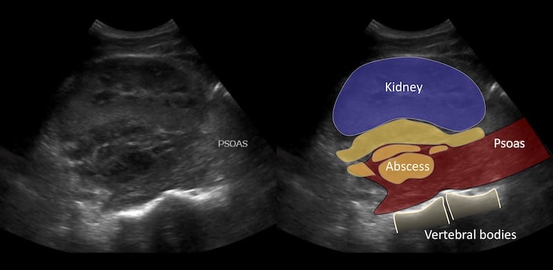
An 18 year old man presents with fevers, right loin pain and dysuria. He has pyuria. You wonder about pyelonephritis or even an infected obstructed kidney so take a look with ultrasound.
Describe and interpret these scans
IMAGE INTERPRETATION
CLINICAL CORRELATION
Renal abscess with retroperitoneal extension and psoas involvement
Psoas abscess is a rare pathology. It may occur from haematogenous or lymphatic spread, contiguous extension from adjacent infection or occur from traumatic inoculation from penetrating injury. In this case the abscess was extensive involving the renal parenchyma, the retroperitoneal space between the kidney and psoas and the psoas muscle itself. It was not clear whether the origin of the abscess was the kidney with spread to psoas, or the psoas with spread to the kidney. The organism grown both from urine and percutaneous image guided aspiration of the psoas collection grew Escherichia coli perhaps favouring direct extension from the kidney.
Further reading
- Bell AE. Psoas Abscess. 5-Minute Clinical Consult
[cite]
TOP 100 ULTRASOUND CASES
An Emergency physician based in Perth, Western Australia. Professionally my passion lies in integrating advanced diagnostic and procedural ultrasound into clinical assessment and management of the undifferentiated patient. Sharing hard fought knowledge with innovative educational techniques to ensure knowledge translation and dissemination is my goal. Family, wild coastlines, native forests, and tinkering in the shed fills the rest of my contented time. | SonoCPD | Ultrasound library | Top 100 | @thesonocave |


