![]()
Ultrasound Case 090
Presentation
A 30 year old woman presents with right iliac fossa pain. Quantitative BHCG is positive at 2500 IU. Her bladder is empty so you proceed to transvaginal ultrasound.
View 2: Uterus longitudinal.

View 3: Right ovary in transverse
View 4: Right ovary in transverse with colour Doppler
View 5: Gestation sac, transverse and longitudinal views

Describe and interpret these scans
IMAGE INTERPRETATION
Image 1: Uterus transverse. This shows the uterus in transverse section, with more echogenic endometrium centrally. Embedded within that is a hypoechoic gestation sac. Within the tiny sac is a small echogenic focus (12 o’clock) that will develop into the yolk sac.
Image 2: Uterus longitudinal. The gestation sac is seen in the longitudinal plane.
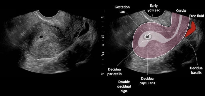
Image 3: Right ovary in transverse. This was the site of maximal tenderness. The probe is gently pushed and the corpus luteum which is part of the ovary is seen to move with the ovary.
Image 4: Right ovary in transverse with colour Doppler. The hyperaemia surrounding a corpus luteum cyst is characteristic and known as a “Ring of Fire”. An ectopic often has the same peripheral hyperaemia.
Image 5: Gestation sac in transverse and longitudinal images. The average diameter is measured and calculated. It is then compared to standard reference values and gestational age determined. At 4mm gestational age is about 4 +4/40 (4 weeks and 4 days). The sac grows an average of 1mm per day.
CLINICAL CORRELATION
Early intrauterine pregnancy and right sided corpus luteum causing pain.
Is it an IUP?
Early in gestation it is very difficult to be certain a small hypoechoic cystic structure within the endometrium is an early sac. The presence of a definite yolk sac or embryo are absolute evidence. Before this several features can strongly suggest this is an early sac, but are not completely definitive. These features include that the IUP is spherical and buried within the endometrium, and is surrounded by an echogenic ring – the decidual capsularis, then a slightly hypoechoic layer, then the next echogenic surrounding layer, the decidua parietalis. This is known as the double decidual sign. In this case the very early yolk sac is visualised but it is so small it looks more like a nodule than a sac.
Is it a corpus luteum or an ectopic?
The donut appearance of a tubal ectopic sitting next to the ovary can appear very similar to the appearance of a corpus luteum. Both may have a hypoechoic centre and echogenic surround. Both are hyperaemic when interrogated by colour Doppler. The best way to tell is to assess dynamically. Position the probe against the ovary / donut shaped mass and gently press. If the object of interest is a CL cyst and is part of the ovary they will move together – as in this case. If the object is separate from the ovary they will slide against each other or separate (see Top 100 Case 017).
[cite]
TOP 100 ULTRASOUND CASES

An Emergency physician based in Perth, Western Australia. Professionally my passion lies in integrating advanced diagnostic and procedural ultrasound into clinical assessment and management of the undifferentiated patient. Sharing hard fought knowledge with innovative educational techniques to ensure knowledge translation and dissemination is my goal. Family, wild coastlines, native forests, and tinkering in the shed fills the rest of my contented time. | SonoCPD | Ultrasound library | Top 100 | @thesonocave |