
![]()
FFS: Cerebral haemorrhage
This article addresses cerebral haemorrhage, particularly spontaneous intracerebral haemorrhage (ICH), which has the highest morbidity and mortality of all stroke types.
- CT brain ± CT angiogram is the key urgent imaging in ED.
Stroke refers to a vascular event causing a focal neurological deficit lasting >24 hours. Use of specific terms—cerebral infarction or cerebral haemorrhage—is preferred, as these represent distinct entities with different management.
Pathophysiology
Causes of cerebral haemorrhage:
- Hypertension:
- Common locations:
- Basal ganglia
- Cerebellum
- Brainstem: midbrain, pons, medulla
- Haemorrhage outside these typical regions suggests alternative pathology.
- Common locations:
- Amyloid angiopathy:
- Seen in elderly; lobar distribution
- Subarachnoid haemorrhage (SAH)
- Secondary causes:
- Trauma
- Coagulopathy
- Tumour
- AV malformation
- Infection
- Vasculitis
Complications
- Raised intracranial pressure: ↓ consciousness, airway compromise
- Intraventricular extension → hydrocephalus
- Seizures
- Aspiration pneumonia
- Long-term neurological deficits
Clinical assessment
History clues suggesting haemorrhage:
- Sudden headache, nausea/vomiting
- Altered consciousness
- Seizures
- Sudden collapse
Medications: warfarin, DOACs
Comorbidities and baseline function impact management.
Examination essentials:
- Airway, breathing, circulation; check gag reflex
- Blood glucose
- Neurological status: GCS, localising signs
Stroke syndromes (less distinct in haemorrhage than infarction):
- Anterior circulation (carotid):
- ACA: contralateral leg weakness
- MCA: face/arm > leg weakness, aphasia, neglect, gaze deviation
- Posterior circulation (vertebrobasilar):
- PCA: visual disturbances, cortical blindness
- Brainstem/cerebellum: vertigo, diplopia, dysarthria, ataxia, crossed signs
Stroke severity scoring:
- Use NIHSS (formal), FAST (initial screening)
Assessment of severity
ICH Score (predicts mortality):
- GCS
- Age ≥ 80
- ICH volume ≥ 30 mL (via ABC/2 method)
- Intraventricular extension
- Location: supratentorial vs infratentorial
See related documents: ICH Score, Volume Estimation.
Differential diagnoses
- Tumours
- Subdural/extradural haematoma
- Venous sinus thrombosis
- Metabolic: hypo/hyperglycaemia, hyponatraemia, Wernicke’s
- Todd’s paresis
- Demyelination
- Functional disorders
Investigations
Blood tests:
- FBC
- U&Es / glucose
- Coagulation profile (especially for anticoagulant use)
- ESR/CRP (vasculitis)
ECG: may show nonspecific changes
CXR: check for aspiration in decreased GCS
CT brain (non-contrast): essential for all suspected strokes
- Rules out:
- ICH, SAH, extra-axial bleeds
- Stroke mimics (tumour, venous thrombosis)
Typical hypertensive bleed sites:
- Brainstem
- Cerebellum
- Basal ganglia
Signs of raised ICP:
- Sulcal effacement
- Midline shift
- Ventricular compression
- Quadrigeminal cistern obliteration (“smiley face”)
CT angiogram:
- Detects:
- Vascular malformations (AVMs, aneurysms)
- Ongoing bleeding (“spot sign”)
- Active contrast leak into haematoma on CTA
- Predicts haematoma expansion and poor outcome
- On non-contrast CT, shows active bleeding (unclotted blood)
Scan urgency:
Prioritise if:
- Thrombolysis/thrombectomy candidate
- Suspected SAH
- ↓ GCS
- Coagulopathy or anticoagulant use
MRI/A:
- MRI may clarify diagnosis if CT inconclusive
- MRA useful for underlying lesions
Management
General approach:
- ABC resuscitation
- Supplemental O₂ if saturations < 95%
- IV access and fluids (avoid glucose)
- Nil orally pending swallowing assessment
- Anticoagulation reversal:
- Warfarin: Vit K + prothrombinex + FFP
- DOACs: idarucizumab (dabigatran), andexanet alfa (Xa inhibitors)
- Glucose management:
- Avoid both hypo- and hyperglycaemia
- IDC for reduced GCS or nursing needs
- Hypertension control:
- Target SBP ≈140 mmHg
- IV options: hydralazine, GTN, esmolol, labetalol, nicardipine
- Antiplatelet reversal:
- Do not give platelets unless active bleeding or procedure
- Pyrexia: treat if T > 37.5°C
Specific considerations
SAH:
- Urgent neurosurgical referral
Posterior fossa haemorrhage:
- Cerebellar bleed >3 cm = urgent surgical decompression
- Brainstem haemorrhage = poor prognosis; discuss with neurosurgery
Supratentorial ICH:
- Surgery not routinely beneficial
- Discuss with neurosurgery if:
- Young patient, good premorbid state
- Hydrocephalus
- Mass effect
- Presenting <24 hrs
Decision factors:
- Age < 80
- Baseline function
- GCS 5–15 or severe focal deficit
- CT findings
- Family expectations
- Likelihood of survival and recovery
Surgery should occur within 72 hours if indicated.
Disposition
- SAH or posterior fossa ICH: urgent transfer to neurosurgical centre
- Supratentorial ICH: transfer if surgery indicated
- Others: admit to stroke unit
Plan for possible deterioration. Decisions regarding treatment limitations should be made early, especially for high-risk patients.
In younger patients, consider early transfer to neurosurgical facility if deterioration may require surgical intervention.
Appendix 1
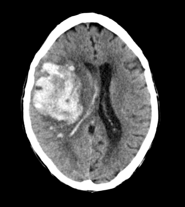
Large intracerebral haemorrhage in the region of the basal ganglia – a typical – “hypertensive” site for bleeding. There is significant mass effect with midline shift, effacement of sulci and obliteration of the right lateral ventricle.
Appendix 2
Intracerebral Haemorrhage Volume Estimation:
Calculation Method:
Estimated volume of an ellipsoid lesion = A x B x C / 2
- A = greatest haemorrhage diameter in the axial plane
- B = haemorrhage diameter at 90º to A in the axial plane
- C = originally described as the number of CT slices with haemorrhage multiplied by the slice thickness, but can simply be substituted with the cranio-caudal diameter of the haemorrhage where there is access to multiplanar reformats
If the measurements are made in centimetres (cm), then the volume will be in cubic centimetres (cc). Note that 1 cm3 = 1 ml
Interpretation:
- An ICH volume of ≥ 30ml is one of 5 parameters thatpredicts a more serious hemorrhage within the Intracerebral Hemorrhage (ICH) Score
- A baseline intracerebral haemorrhage volume of > 50 – 60 ml is a poor prognostic marker.
Appendix 3
The Intracerebral Haemorrhage (ICH) Score
Scoring method:
| Parameter | Result | Score |
|---|---|---|
| Glasgow Coma Score (GCS) | 13–15 | 0 |
| 5–12 | 1 | |
| 3–4 | 2 | |
| Age ≥ 80 | No | 0 |
| Yes | 1 | |
| ICH volume ≥ 30 mL | No | 0 |
| Yes | 1 | |
| Intraventricular haemorrhage | No | 0 |
| Yes | 1 | |
| Origin of haemorrhage | Supratentorial | 0 |
| Infratentorial | 1 |
Interpretation:
| ICH Score | 30-day mortality |
|---|---|
| 0 | 0% |
| 1 | 13% |
| 2 | 26% |
| 3 | 72% |
| 4 | 94% |
| 5 | 100% |
| 6 | 100% |
References
Publications
- Sharma R. CT angiographic spot sign (intracerebral haemorrhage). Radiopaedia
- Kusel K. Swirl sign (intracranial haemorrhage). Radiopaedia
- Clinical Guidelines for Acute Stroke Management – National Stroke Foundation
- Brazis PW, Masdeu JC, Biller J. Localization in Clinical Neurology. 8e 2021
- Fuller G. Neurological Examination Made Easy. 6e 2019
- O’Brien M. Aids to the Examination of the Peripheral Nervous System. 6e 2023
FOAMed
- Gibbs M. Neuroimaging Cases 004. LITFL
- Flower O. EVACUATE: ICH Management. LITFL
- Suresh Kochath P. CT Case 078. LITFL
- Lam L. CT Case 022. LITFL
- Coni R. Neuro 101: Cerebral Hemispheres. LITFL
Fellowship Notes

MBBS DDU (Emergency) CCPU. Adult/Paediatric Emergency Medicine Advanced Trainee in Melbourne, Australia. Special interests in diagnostic and procedural ultrasound, medical education, and ECG interpretation. Co-creator of the LITFL ECG Library. Twitter: @rob_buttner

Educator, magister, munus exemplar, dicata in agro subitis medicina et discrimine cura | FFS |