![]()

CT Case 066
A 71-year-old presents with vomiting and abdominal pain and inability to open his bowels. This was preceded by three days of diarrhoea.

![]()
A 71-year-old presents with vomiting and abdominal pain and inability to open his bowels. This was preceded by three days of diarrhoea.

The Medication Access and Training Expansion (MATE) Act went into effect late 2023. Here is a review of the Top 8 CME Courses to Meet Your Substance Use Disorder Training Requirements
A 70-year-old presents with flank pain 4 days after a renal biopsy for investigation of a renal mass. He has a previous history of bioprosthetic aortic valve and is on warfarin and aspirin.
A 70-year-old presents with flank pain 4 days after a renal biopsy for investigation of a renal mass. He has a previous history of bioprosthetic aortic valve and is on warfarin and aspirin.
A 86-year-old lady tripped over while walking, landing face first on the ground. She has sustained multiple abrasions to her face, an eyelid laceration and she has midline c-spine tenderness

Barrett’s oesophagus: reflux-driven columnar metaplasia of the distal oesophagus and precursor to adenocarcinoma—history, key figures, diagnosis, management
A 65-year-old lady presents with abrupt onset of epigastric pain radiating to her back and right shoulder.
A 36-year-old female presents with sudden onset right sided facial drop, right hemiparesis and dysarthria.
May 2024 Implantable device imaging and interpretation. Ventriculoperitoneal (VP) shunts with Friedman, Patterson and Miller

James Douglas (1675 - 1742) was a Scottish physician and anatomist. Pouch of Douglas; folds of Douglas; and line of Douglas

Network Five Emergency Medicine Journal Club Episode 29 reviewing papers on LGBTQIA+ identity and healthcare!
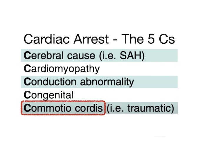
Cardiac Arrest - the 5 C's with Cliff Reid. Causes Of Cardiac Arrest You NEVER Thought Of?! Meet The Hs & Ts & Cs!