![]()

Paget-Schroetter Syndrome
Primary thrombosis of the subclavian vein at the costoclavicular junction. The formation of an axillo-subclavian vein thrombosis results from endothelial trauma, often as a result of repetitive activity of the upper limbs.

![]()
Primary thrombosis of the subclavian vein at the costoclavicular junction. The formation of an axillo-subclavian vein thrombosis results from endothelial trauma, often as a result of repetitive activity of the upper limbs.

Berkeley George Andrew Moynihan, Lord Moynihan of Leeds (1865-1936) was an English General surgeon. Eponymously associated with the Moynihan sign (1905), an adaptation of Murphy's sign, a method used to differentiate pain in the right upper quadrant.

The original hope was that using AI might allow us to create a single podcast episode in one afternoon. But things didn’t go quite the way we planned.
Native Hip Dislocations. Adult Orthopedic case interpretation with Carrie Bissell, Ainsley Bloomer, Aaron Fox, Andrew Rees and Kendrick Lim
AI can work for Spotify's music DJ, so we wondered if it can also work for a cardiology podcast intended for clinicians. Here are the pros and cons we discovered.
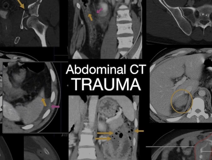
Abdominal CT: bladder injuries. Severe blunt force injuries to the pelvis not only cause fractures but can also injure the urinary bladder.
Abdominal CT: pelvic fractures. We consider three types of pelvic fractures and learn If you see one pelvic fracture, you will find others
Abdominal CT: body wall injuries. Fractures are commonly associated with injury to the abdominal muscles. These injuries can have a wide range of appearances
Abdominal CT: Trauma series. Describing and diagnosing rib fractures - lower ribs are included in an abdominal CT
Abdominal CT: spinal fractures. Trauma patients undergoing imaging should always be evaluated for spinal fractures.
Abdominal CT: Trauma series. Describing and diagnosing diaphragmatic injuries
Abdominal CT: Trauma series. Evaluating bowel and mesenteric trauma with examples cases