


Cope psoas test (ilio-psoas rigidity test; psoas extension test) is a physical examination manoeuvre used to identify irritation of the iliopsoas muscle by an adjacent inflammatory process, classically associated with retrocaecal appendicitis.
With the patient lying on the unaffected side, passive extension of the affected hip may reproduce abdominal or flank pain. The test has low sensitivity, and a negative result does not exclude appendicitis.
Modern examination technique
Two principal manoeuvres are commonly described for eliciting the psoas sign.
Passive hip extension (Cope technique): With the patient lying in the left lateral decubitus position, the examiner stabilises the pelvis and gently extends the right hip by moving the thigh posteriorly. Reproduction of right-sided abdominal or flank pain suggests irritation of the iliopsoas muscle by an adjacent inflammatory process.
Resisted hip flexion: With the patient supine, the examiner asks the patient to raise the right thigh or straight leg against resistance. Reproduction of right-sided abdominal pain is considered a positive response.
The two manoeuvres are frequently grouped under the term psoas sign, although they involve different mechanical actions: passive stretching of the iliopsoas versus active contraction against resistance.
Historical development
1902 – Samuel James Meltzer (1851-1920) described the use of active iliopsoas contraction to assist palpation and diagnosis of structures within the iliac fossa. He instructed patients to raise the extended leg slightly from the bed, producing a palpable muscular elevation beneath the appendix and caecum.
Meltzer observed that active contraction could intensify tenderness in appendicitis, facilitate localisation of the appendix, and help distinguish intra-abdominal inflammation from abdominal wall myositis. He found the technique particularly useful in chronic and subacute cases.
Although predating Cope’s description, Meltzer’s technique combined active iliopsoas contraction with abdominal palpation, whereas Cope used passive hip extension to identify irritation or rigidity of the muscle.
1921 – Sir Vincent Zachary Cope (1881-1974) described the examination for determination of ilio-psoas rigidity in the first edition of The Early Diagnosis of the Acute Abdomen. He recognised that inflammation adjacent to the psoas muscle could cause reflex hip flexion to relieve pain. Lesser degrees of irritation could be demonstrated by placing the patient on the unaffected side and passively extending the affected thigh.
It is well known that if there be an inflamed focus in relation to the psoas muscle the corresponding thigh is often flexed by the patient to relieve the pain. A lesser degree of such contraction (and irritation) can be determined often by making the patient lie on the opposite side and extending the thigh on the affected side to the full extent. Pain will be caused by the maneuver if the psoas is rigid from either reflex or direct irritation (Fig 6.). The value of the test is diminished if the abdominal wall be rigid. The psoas-test is not so easily elicited when the inflammation becomes subacute.
Cope 1921
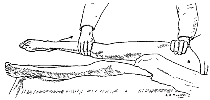
1929 – Hamilton Bailey included the psoas test among three clinical signs useful in difficult cases of suspected appendicitis. He instructed the examiner to place the patient on the left side, fully extend the hip and abduct the thigh. Pain during this manoeuvre suggested irritation of the psoas by an adjacent inflamed appendix.
Diagnostic accuracy
2018 – Rastogi et al reviewed the historical descriptions and diagnostic performance of abdominal examination signs, reporting a sensitivity of 13–42% and specificity of 79–97% for the psoas sign in acute appendicitis. They cited Andersson’s 2004 Meta-analysis of the clinical and laboratory diagnosis of appendicitis, which reported a positive likelihood ratio of 2.31 and negative likelihood ratio of 0.85.
2022 – Yale, Tekiner and Yale highlighted the limited evidence supporting the psoas, obturator and Rovsing signs. Many studies failed to specify the examination technique, standardise performance or correlate findings with the anatomical position of the appendix.
The published estimates cannot reliably be attributed to Cope’s original passive hip-extension manoeuvre. A negative psoas test does not exclude appendicitis, and the modest positive likelihood ratio limits its value as an isolated diagnostic finding.
Eponymythology
Cope’s original ilio-psoas rigidity test involved passive extension of the affected thigh with the patient lying on the opposite side. Hamilton Bailey described an additional component of hip abduction in 1929.
| Feature | Cope, 1921 | Bailey, 1929 | Contemporary variant |
|---|---|---|---|
| Position | Opposite lateral decubitus | Left lateral decubitus | Often supine |
| Action | Passive hip extension | Hip extension and abduction | Active resisted hip flexion |
| Mechanism | Stretch of irritated iliopsoas | Stretch of irritated iliopsoas | Contraction of iliopsoas |
| Positive finding | Pain on extension | Pain on movement | Right-sided abdominal pain |
Modern descriptions frequently include active resisted hip flexion as an alternative means of eliciting the psoas sign. Although both manoeuvres assess irritation of the iliopsoas muscle, they involve different mechanical actions: passive stretching versus active contraction.
Published diagnostic accuracy studies rarely distinguish these techniques, limiting the applicability of their findings to Cope’s original examination.
Associated Persons
- Sir Vincent Zachary Cope (1881-1974)
Alternative names
- Ilio-psoas test
- iliopsoas rigidity, extension test
- ilio-psoas rigidity test
- Obraztsov symptom
References
Historical references
- Meltzer SJ. On the contraction of the iliopsoas muscle as an aid in the diagnosis of the contents of the iliac fossa. New York Medical Journal. 1902 July 19;76:100–103.
- Cope VZ. The early diagnosis of the acute abdomen, 1921: 43-44
- Bailey H. Physical signs in acute abdominal catastrophies. Medical review of reviews 1929; 35: 161-170
Eponymous term review
- Butsenko V. N., Antoniuk S. M. [Significance of the separate symptoms in acute appendicitis]. Klinicheskaia khirurgiia Klin. Khir. 1992;(2):33-5
- Augustin G. Acute Appendicitis. In: Acute Abdomen During Pregnancy. Springer International Publishing; 2014:3-43
- Berry J Jr, Malt RA. Appendicitis near its centenary. Ann Surg. 1984 Nov;200(5):567-75.
- Izbicki JR, Knoefel WT, Wilker DK, Mandelkow HK, Müller K, Siebeck M, Schweiberer L. Accurate diagnosis of acute appendicitis: a retrospective and prospective analysis of 686 patients. Eur J Surg. 1992 Apr;158(4):227-31
- Andersson RE. Meta-analysis of the clinical and laboratory diagnosis of appendicitis. Br J Surg 2004; 91(1): 28-37
- Rastogi V, Singh D, Tekiner H, Ye F, Mazza JJ, Yale SH. Abdominal Physical Signs and Medical Eponyms: Movements and Compression. Clin Med Res. 2018 Dec;16(3-4):76-82.
- Moris D, Paulson EK, Pappas TN. Diagnosis and Management of Acute Appendicitis in Adults: A Review. JAMA. 2021;326(22):2299–2311.
- Yale SH, Tekiner H, Yale ES. Signs and syndromes in acute appendicitis: A pathophysiologic approach. World J Gastrointest Surg. 2022 Jul 27;14(7):727-730.
- Yale SH, Tekiner H, Yale ES. Utility of bedside signs in diagnosing acute appendicitis: a neurophysiological perspective. Pediatr Surg Int. 2026 Mar 6;42(1):142.
eponymictionary
the names behind the name

BA MA (Oxon) MBChB (Edin) FACEM FFSEM. Emergency physician, Sir Charles Gairdner Hospital. Passion for rugby, medical history, medical education, and asynchronous learning #FOAMed evangelist. Editor-in-Chief and Webmaster of Life in the Fast lane, LITFL | On Call 4e| Eponyms | Books | Horology |