![]()
CT Case 091
A 40-year-old female, presents with headache and abrupt left sided visual disturbance. She has a history of T2DM and hypertension.
On examination she has a left homonymous hemianopia.
There are no other neurological deficits.
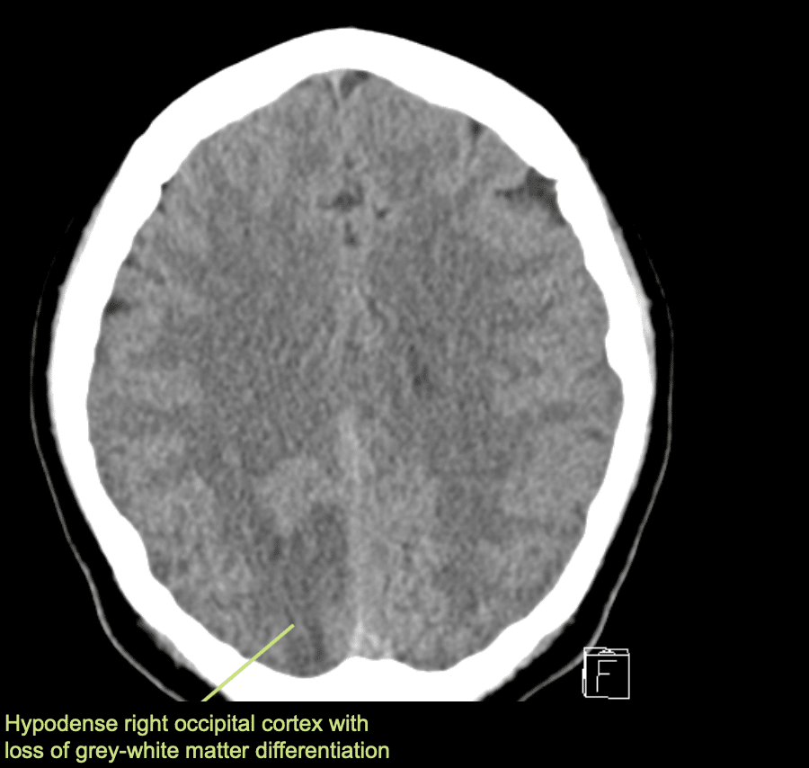
Her CT brain is shown below
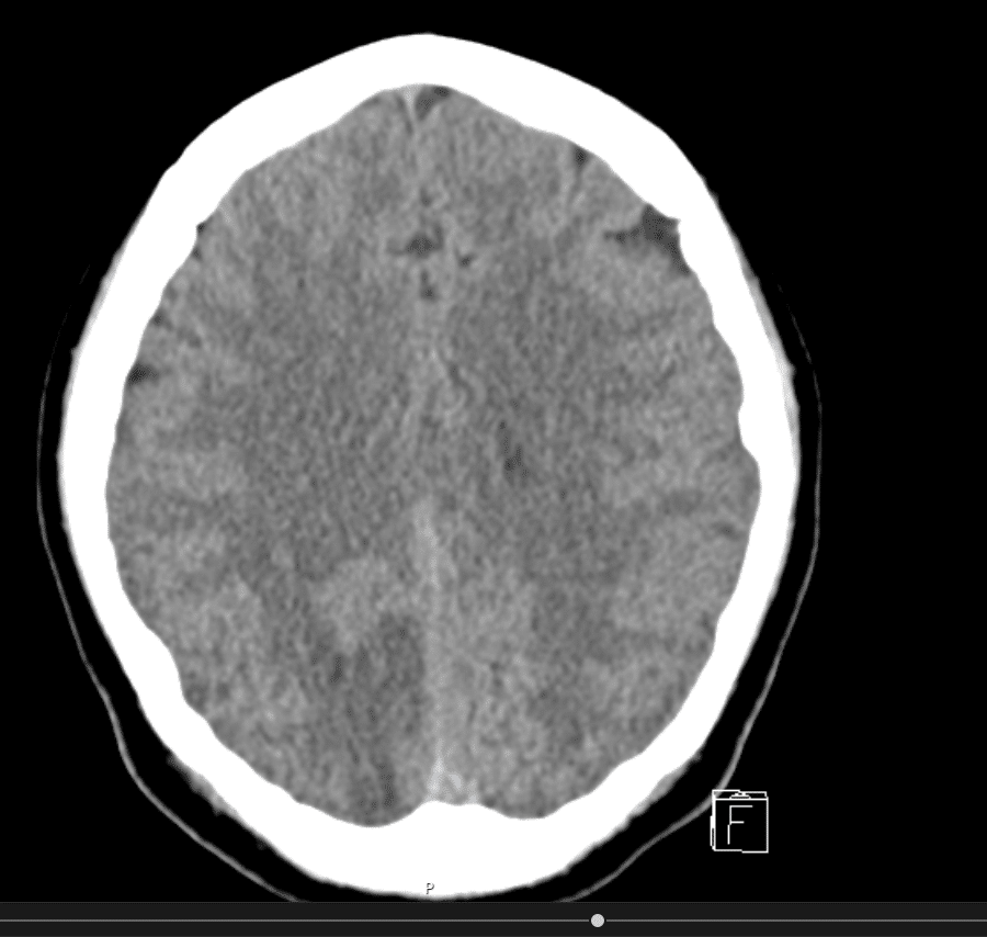
Describe and interpret the CT scans
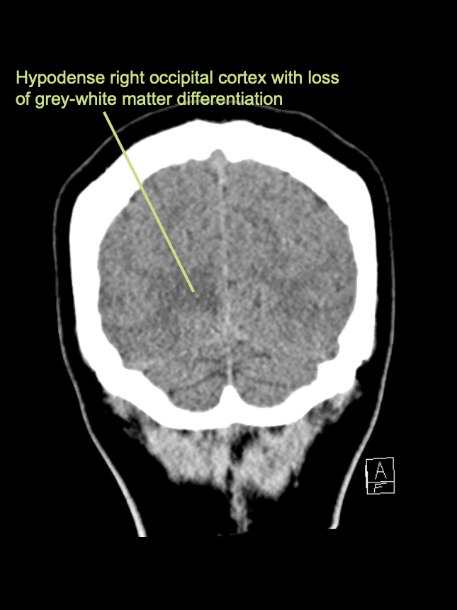
There is an area of hypodensity and loss of grey-white matter differentiation within the right occipital lobe in keeping with a cortical infarct.
This represents a right PCA territory infarct.
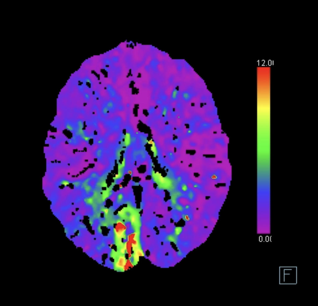
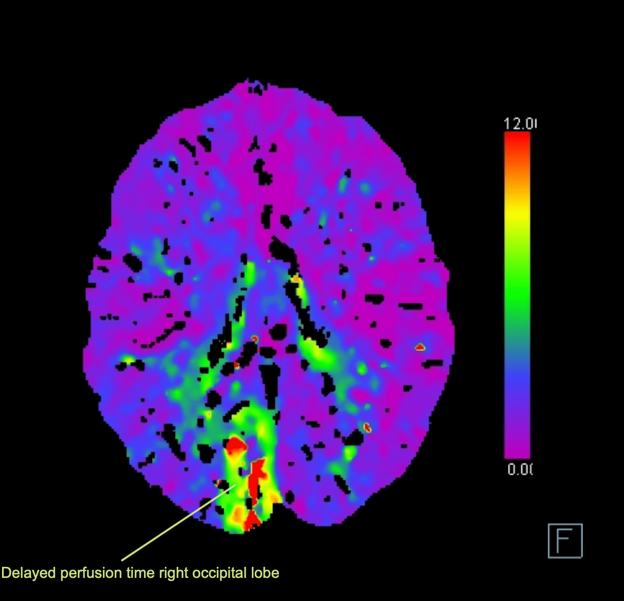
The image from the CT perfusions study shows the Tmax, that is the time delay for the IV contrast to reach the brain parenchyma. We see a delay in brain parenchyma perfusion in the right occipital lobe.
As the CT perfusion studies show a large core and small penumbra, this case wasn’t suitable for thrombolysis.
She was managed with DAPT and optimisation of her blood pressure and blood sugar.
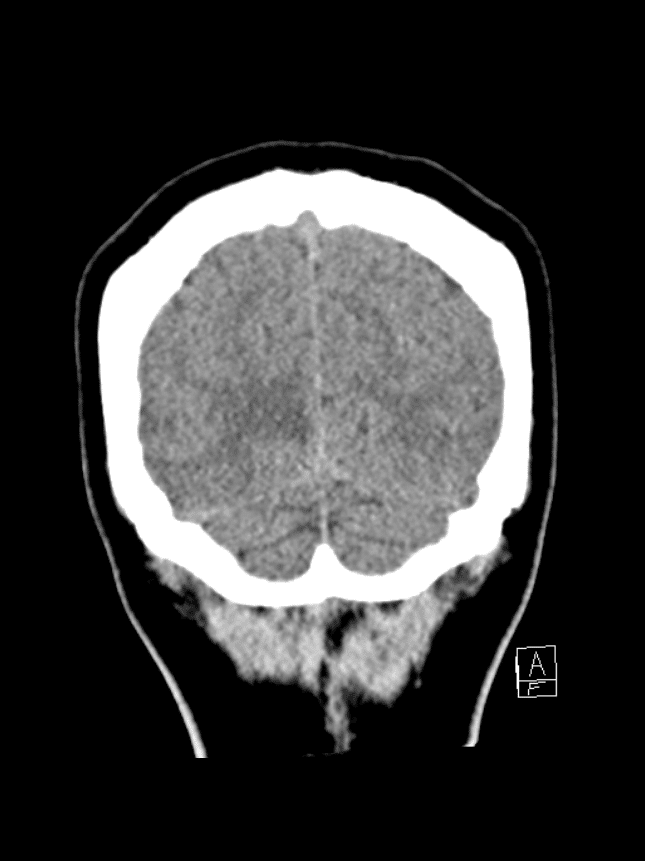
She presents again three years later following a seizure.
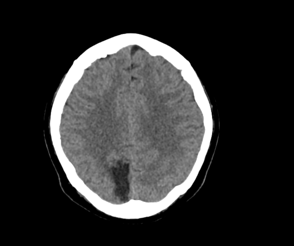
Another CT brain is performed
Describe and interpret the CT scans
There is an established infarct in the right occipital lobe which was determined to be the focus for her seizures and she was commenced on anticonvulsants on discharge.

Clinical Pearls
Occipital strokes represent approximately 10% of all ischaemic strokes.
Patients are more likely to experience a headache compared to those with anterior circulation strokes.
Most commonly patients will present with a combination of headache and visual field defect.
Residual visual field defects can result in significant disability.
Patients with PCA stroke tend to be younger and are more frequently female.
As with all ischaemic strokes the principles of management include thrombolysis or clot retrieval (in the appropriate patient), and subsequently blood pressure and glycaemic management.
5-7% of patients who have had a stroke will subsequently develop epilepsy.
References
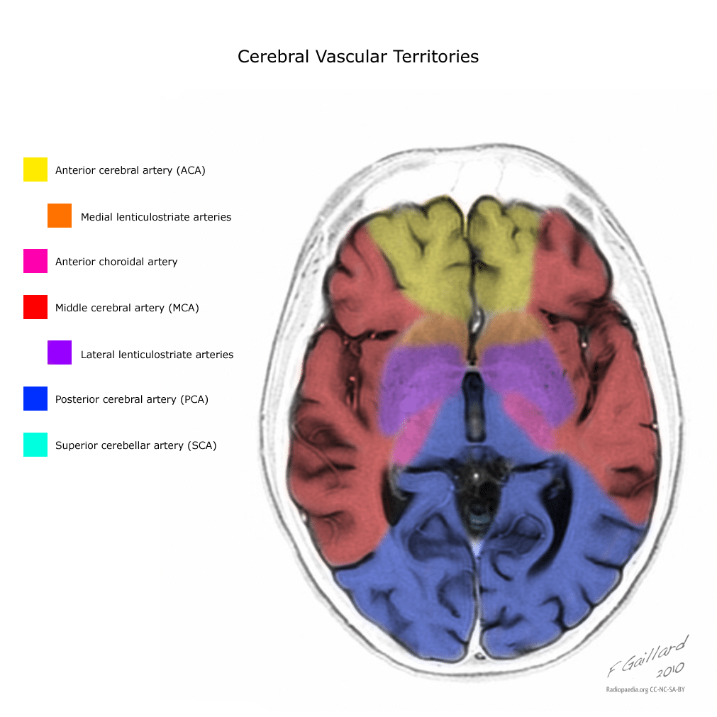
- Galliard F. Cerebral vascular territories (illustration). Radiopaedia
TOP 100 CT SERIES

Emergency Medicine Education Fellow at Liverpool Hospital NSW. MBBS (Hons) Monash University. Interests in indigenous health and medical education. When not in the emergency department, can most likely be found running up some mountain training for the next ultramarathon.

Dr Leon Lam FRANZCR MBBS BSci(Med). Clinical Radiologist and Senior Staff Specialist at Liverpool Hospital, Sydney

Sydney-based Emergency Physician (MBBS, FACEM) working at Liverpool Hospital. Passionate about education, trainees and travel. Special interests include radiology, orthopaedics and trauma. Creator of the Sydney Emergency XRay interpretation day (SEXI).

Provisional fellow in emergency radiology, Liverpool hospital, Sydney. Other areas of interest include paediatric and cardiac imaging.