![]()
CT Case 092
A 92-year-old man presented to emergency with abdominal pain, vomiting and fever. He had been experiencing 24 hours of abdominal pain which had become significantly worse in the last 3 hours.
He has generalised abdominal tenderness and distention.
Lactate is 2.5 and he has a mild AKI.
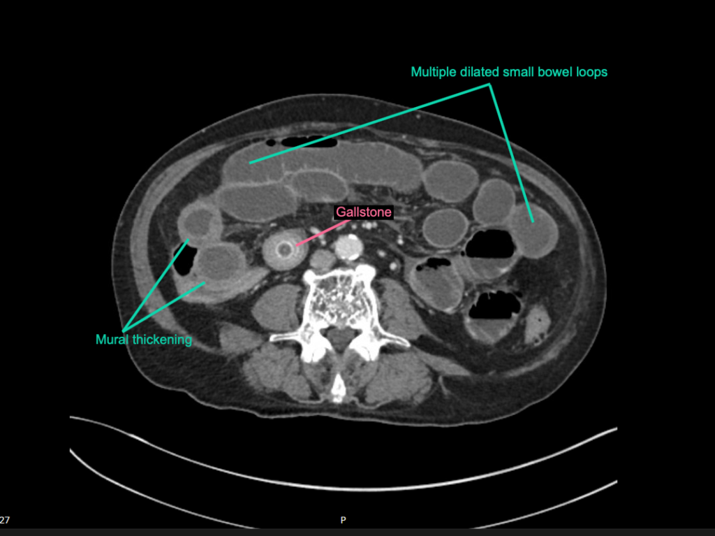
His CT abdomen is shown below
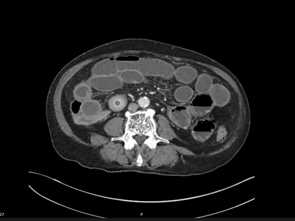
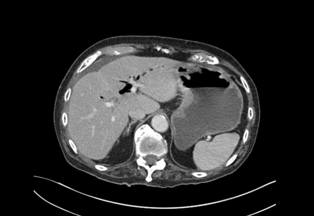
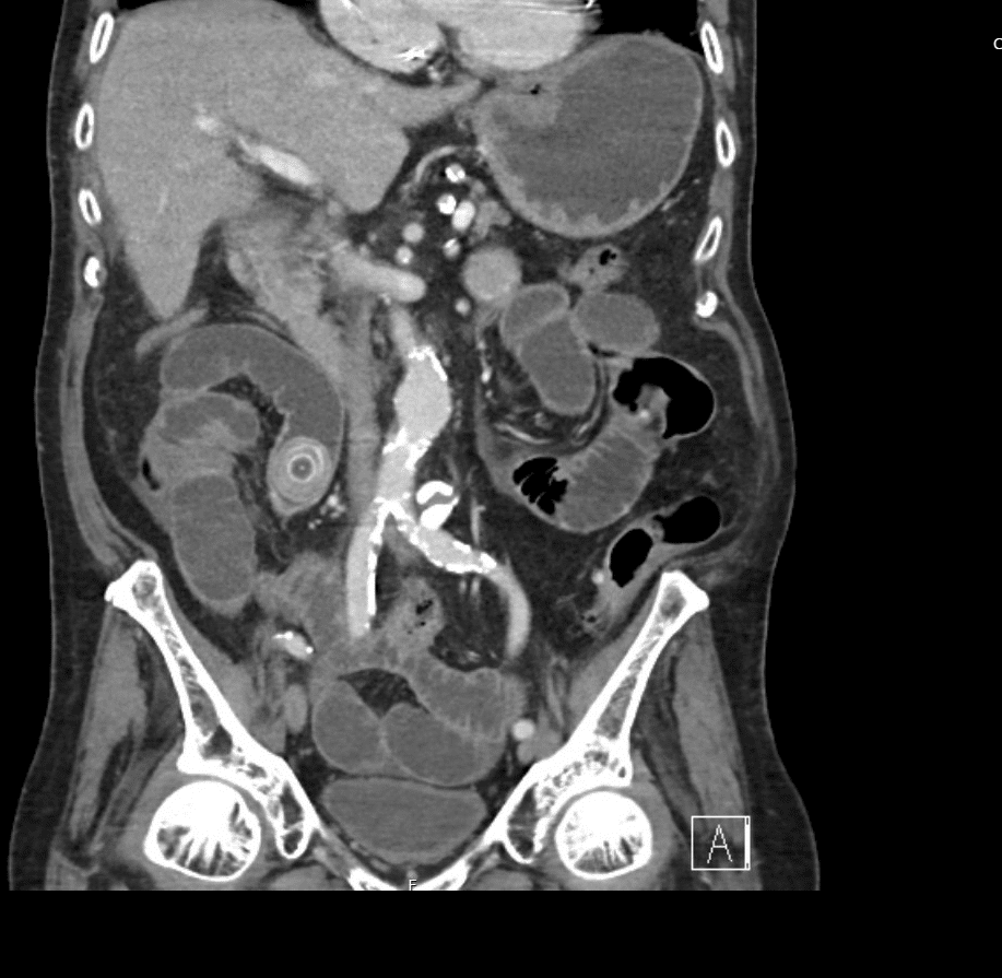
Describe and interpret the CT scans
There are multiple dilated loops of small bowel secondary to small bowel obstruction. There is mild mural thickening in the right lower quadrant, suggestive of early ischaemia.
An abrupt transition is seen in the terminal ileum due to a 30 mm gallstone – a gallstone ileus.
Pneumobilia is present, this is secondary to a cholecystoduodenal fistula.


Clinical Pearls
Gallstone ileus is an uncommon but potentially serious cause of small bowel obstruction.
Gallstone ileus was first described by Thomas Bartholin (1616-1680) in 1654 (father of Caspar Bartholin the Younger (1655-1738) of the Bartholin cyst).
It occurs when a gallstone enters the small bowel via a biliary-enteric fistula, causing a mechanical bowel obstruction. It is more common in elderly populations with a higher incidence in women.
Symptoms tend to occur over several days due to passage of the stone through the bowel lumen before impaction. The site of obstruction is the terminal ileum in 50-70% cases (as in this case), the proximal ileum and jejunum in 20-40% cases and the duodenum in <10%. Small stones tend to pass spontaneously with larger stones more likely to cause obstruction, 90% of stones over 2cm in diameter cause obstruction. Impacted stones manifest typically with classic signs and symptoms of bowel obstruction. There may be colicky, often periumbilical, abdominal pain, distension, constipation, nausea and vomiting.
The main therapeutic goal is relief of intestinal obstruction by extraction of the gallstone.
Current surgical options are simple enterolithotomy (as in this case); enterolithotomy, cholecystectomy and fistula closure (one-stage procedure) or enterolithotomy with cholecystectomy performed later as a two-stage procedure. Enterolithotomy is the most common surgical procedure performed.
This patient went for urgent laparotomy. Findings were of small bowel obstruction with only minor congestion. A 3cm gallstone was found in the distal ileum. The gallstone was able to be moved proximally, and an enterotomy was performed for removal of the gallstone via the ileum.
References
- Nuño-Guzmán CM, Marín-Contreras ME, Figueroa-Sánchez M, Corona JL. Gallstone ileus, clinical presentation, diagnostic and treatment approach. World J Gastrointest Surg. 2016 Jan 27;8(1):65-76.
- Ayantunde AA, Agrawal A. Gallstone ileus: diagnosis and management. World J Surg. 2007 Jun;31(6):1292-7.
- Markogiannakis H, Messaris E, Dardamanis D, Pararas N, Tzertzemelis D, Giannopoulos P, Larentzakis A, Lagoudianakis E, Manouras A, Bramis I. Acute mechanical bowel obstruction: clinical presentation, etiology, management and outcome. World J Gastroenterol. 2007 Jan 21;13(3):432-7.
- Cadogan M. Rigler triad. LITFL
- Cadogan M. Bouveret syndrome. LITFL
- Lassandro F, Romano S, Ragozzino A, Rossi G, Valente T, Ferrara I, Romano L, Grassi R. Role of helical CT in diagnosis of gallstone ileus and related conditions. AJR Am J Roentgenol. 2005 Nov;185(5):1159-65
- Rippy J. Ultrasound Case 032. LITFL
- Bickle I. Rigler triad (gallstone ileus). Radiopaedia
TOP 100 CT SERIES

Emergency Medicine Education Fellow at Liverpool Hospital NSW. MBBS (Hons) Monash University. Interests in indigenous health and medical education. When not in the emergency department, can most likely be found running up some mountain training for the next ultramarathon.

Dr Leon Lam FRANZCR MBBS BSci(Med). Clinical Radiologist and Senior Staff Specialist at Liverpool Hospital, Sydney

Sydney-based Emergency Physician (MBBS, FACEM) working at Liverpool Hospital. Passionate about education, trainees and travel. Special interests include radiology, orthopaedics and trauma. Creator of the Sydney Emergency XRay interpretation day (SEXI).

Provisional fellow in emergency radiology, Liverpool hospital, Sydney. Other areas of interest include paediatric and cardiac imaging.