
![]()
CXR Case 003
55 yo lady presents with worsening chronic cough and wheeze
Describe and interpret this CXR and CT
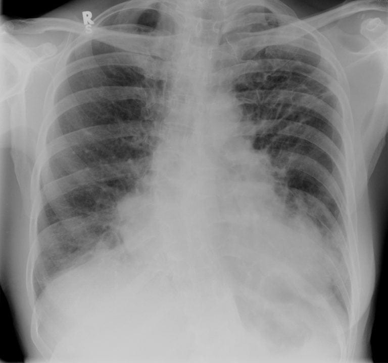
CHEST X-RAY INTERPRETATION
This CXR demonstrates bronchiectasis.
There are coarse, thickened airway markings with ring shadows bilaterally, but worse in the left upper lobe (obscuring the heart border) and left lower lobe, obstructing the hemidiaphragm.
* There is also scoliosis of the spine*
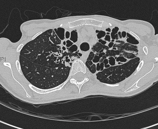
The CT demonstrates bronchiectais with markedly dilated airways and thickened airway walls with patches of sputum plugging.
CLINICAL CORRELATION
Concurrent airways disease is common in bronchiectasis and should be treated similarly to acute asthma
CLINICAL PEARLS
Bronchiectasis patients frequently have airways obstruction which behaves similarly to asthma in presentation and response to treatment.
TOP 150 CXR SERIES
![]()
![]()
![]()
Prof Fraser Brims Curtin Medical School, acute and respiratory medicine specialist, immediate care in sport doc, ex-Royal Navy, academic| Top 100 CXR | Google Scholar | ICIS Course ANZ