
![]()
CXR Case 013
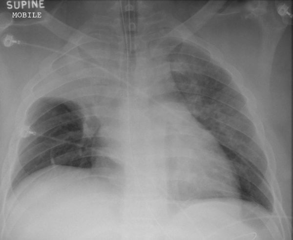
76 year old male presents with severe respiratory distress and is intubated
Describe and interpret this CXR
CHEST X-RAY INTERPRETATION
There is complete collapse of the right upper lobe
* Note the resultant elevation of the right horizontal fissure *
Patchy Left Upper Lobe (LUL) opacification suggestive of consolidation
*ETT and NG tube also present*
CLINICAL CORRELATION
* This is a case of severe pneumonia *
Right upper lobe collapse has distinctive features, and is often easily identifiable on CXR.
Features include: increased density in the right upper lung field, elevation of both the right hilum and right horizontal fissure, and loss of the right cardiomediastinal contour.
CLINICAL PEARLS
A common cause of upper lobe collapse is a proximal tumour or mediastinal mass.
* An acute collapse normally correlates with a sudden worsening of symptoms, particularly breathlessness *
Consider malignancy in patients who present with weight loss, cough and/or haemoptysis
TOP 150 CXR SERIES
![]()
![]()
![]()
Prof Fraser Brims Curtin Medical School, acute and respiratory medicine specialist, immediate care in sport doc, ex-Royal Navy, academic| Top 100 CXR | Google Scholar | ICIS Course ANZ