
![]()
CXR Case 016
86 year old female presents with known bronchiectasis presents with shortness of breath and a worsening cough. She is more hypoxaemic than her normal baseline.
Presenting X-ray 
Repeat X-ray
Describe and interpret this CXR
CHEST X-RAY INTERPRETATION
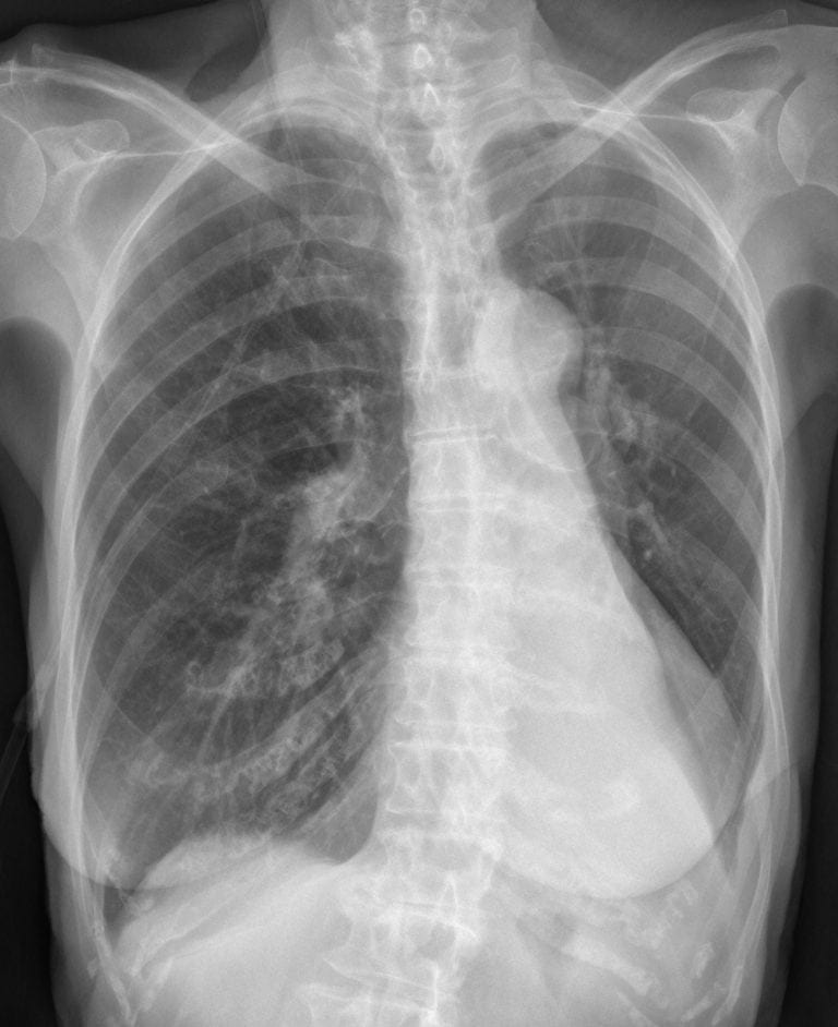
Presenting CXR:
There is complete collapse of the left lower lobe (LLL) creating the sail sign behind the heart and volume loss in the left hemithorax.
There are age-related bony changes of the thoracic vertebral column and ribs
* The lung fields are overinflated, consistent with gas trapping from airways disease *
Presenting CXR:
The left lower lobe is now re-inflated
CLINICAL CORRELATION
Left lower lobe collapse has distinctive features, but may sometimes be missed on CXR.
* Features of left lower lobe collapse include: edge of collapsed lung creating a ‘double cardiac contour,’ loss of normal left hemidiaphragm outline*
A lateral CXR is useful in looking for left lower lobe collapse – a triangular outline representing the collapsed lung may be visible posteriorly.
In this case the patient was hydrated and had lots of chest physio – this is frequently enough to re-inflate collapsed lobes from retained secretions or sputum plugs.
CLINICAL PEARLS
Cardiomegaly may make it difficult to identify left lower lobe collapse.
TOP 150 CXR SERIES
![]()
![]()
![]()
Prof Fraser Brims Curtin Medical School, acute and respiratory medicine specialist, immediate care in sport doc, ex-Royal Navy, academic| Top 100 CXR | Google Scholar | ICIS Course ANZ
Lytic lesions of the lower ribs and increased calcification of the trachea. ? mutliple myeloma