
![]()
CXR Case 035
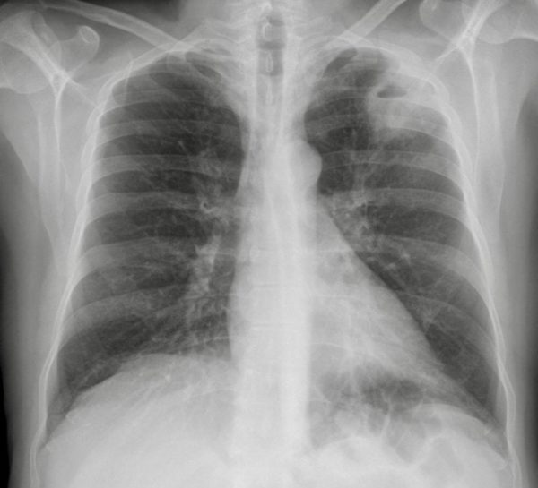
A 21 yo man living in sheltered accommodation presents with fever, malaise and productive cough. He has a history of IV drug use, but has not used within the last 4 weeks. Blood tests reveal raised inflammatory markers.
Describe and interpret this CXR
CHEST X-RAY INTERPRETATION
There is a left upper lobe thick walled cavity with an air fluid level and surrounding consolidation.
*There may be left hilar adenopathy. The rest of the lung field and pleura look normal
CLINICAL CORRELATION
The acute presentation favors infection and abscess, or more unlikely infarct.
Cancer is unlikely given his age and more disseminated infection, e.g. septic emboli is unlikely as there are no other lesions.
TB should be considered given anatomical position, social factors and possibility of immune suppression.
*The hilar adenopathy is likely to be reactive from infection/inflammation.
CLINICAL PEARLS
A CT will help localize the lesion to the anatomical lobe.
A clever chest physio may then be able to lie and tip the patient in the appropriate posture to drain a lung abscess – also providing the all important sputum cultures!
Usually prolonged IV antibiotics are required, rarely surgery.
TOP 150 CXR SERIES
![]()
![]()
![]()
Prof Fraser Brims Curtin Medical School, acute and respiratory medicine specialist, immediate care in sport doc, ex-Royal Navy, academic| Top 100 CXR | Google Scholar | ICIS Course ANZ