
![]()
CXR Case 053
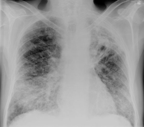
A 79 year old man is placed on non invasive ventilation for progressive type II respiratory failure with a history of worsening cough and fevers.
Describe and interpret this CXR
CHEST X-RAY INTERPRETATION
There is volume loss and fibrosis in the left upper lobe (hilum pulled up).
There is airspace opacification in the right lower lobe on a background of possible fibrosis throughout both lung fields.
CLINICAL CORRELATION
This man has acute right sided pneumonia, exacerbating his already weak ventilatory reserve.
* The left upper lobe fibrosis is highly likely to be caused by TB
CLINICAL PEARLS
*Sending sputum cultures for AFB is mandatory in this situation – this guy had reactivation of his TB – nothing like some positive pressure to help generate aerosolized mycobacterium!
TOP 150 CXR SERIES
![]()
![]()
![]()
Prof Fraser Brims Curtin Medical School, acute and respiratory medicine specialist, immediate care in sport doc, ex-Royal Navy, academic| Top 100 CXR | Google Scholar | ICIS Course ANZ