
![]()
CXR Case 074
A 79 year old male presents with haemoptysis and weight loss over 2 months

click images to enlarge
Describe and interpret this CXR
CHEST X-RAY INTERPRETATION
There is a large mass in the medial right lung adjacent to the mediastinum.
The airway and right inferior pulmonary artery are clearly visualized, suggesting this is posterior to the hilum.
Lung fields, pleura and bones otherwise normal.
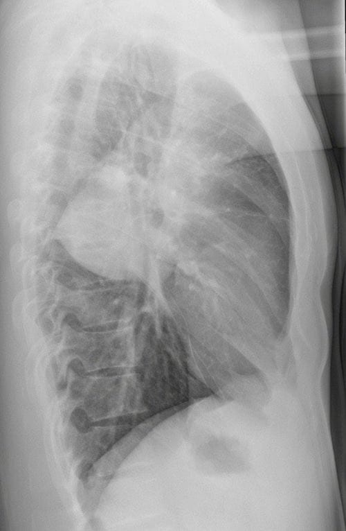
Lateral X-ray confirms the presence of a large mass, posterior to the hilum.
CLINICAL CORRELATION
This is very likely to be a primary lung cancer.
Smoking history, occupational exposures (particularly asbestos) and additional clinical features such as dyspnoea and weight loss should be gained.
CLINICAL PEARLS
Haemoptysis occurs in at least 1 in 5 cases of non small cell lung cancer.
In the majority of cases it is relatively minor and can be managed by stopping any anti-platelet and/or anticoagulants and monitoring closely.
Occasionally tranexamic acid is useful.
TOP 150 CXR SERIES
![]()
![]()
![]()
Prof Fraser Brims Curtin Medical School, acute and respiratory medicine specialist, immediate care in sport doc, ex-Royal Navy, academic| Top 100 CXR | Google Scholar | ICIS Course ANZ