
![]()
CXR Case 117
A 71 yo man with known previous COPD presents with 3 days of worsening cough, fevers and dyspnoea.
click images to enlarge
Describe and interpret this CXR and CT chest
IMAGE INTERPRETATION
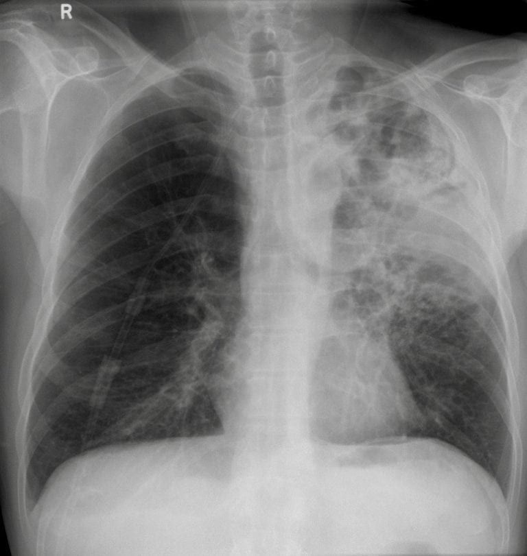
CXR Interpretation:
There is patchy airspace shadowing in the left upper lobe, more dense inferiorly.
There is some volume loss on the left and multiple rounded areas representing non-consolidated emphysema.
Remaining parenchyma and pleura are normal.
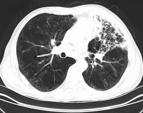
CT Chest Interpretation:
CT chest confirms the presence of emphysema with surrounding consolidation.
CLINICAL CORRELATION
Intercurrent infection is very common in COPD.
The appearances on CXR are sometimes thought to be abscesses – but there is no fluid level and there are just too many to make this likely.
CLINICAL PEARLS
This man needs a repeat CXR in ~8 weeks to ensure that there is no lung cancer within the area of consolidation (the initial CT chest will not reliably tell us this).
TOP 150 CXR SERIES
Prof Fraser Brims Curtin Medical School, acute and respiratory medicine specialist, immediate care in sport doc, ex-Royal Navy, academic| Top 100 CXR | Google Scholar | ICIS Course ANZ