![]()
David Spodick
David H. Spodick (1927-2019) was an American Cardiologist.
Clinician, researcher, educator, and administrator in cardiovascular medicine for over 50 years. World renowned for his work in pericardial diseases and electrocardiology research, diseases of the atria and interatrial conduction abnormalities and cardiovascular physical examination
Spodick, a pioneer in the study of the pericardium, described the classic four-stage evolution of electrocardiographic changes with pericarditis, including ST elevations and PR depressions in 1973/1974. He is eponymously affiliated with Spodick sign in pericarditis (1974) and the Spodick sign of aortic stenosis (1976)
…it is remarkable that clavicular transmission of aortic murmurs seems to have been neglected, while auscultation over the carotid artery has become a clinical ritual. Yet murmurs are nearly always greatly attenuated over the carotid artery, even to the point of disappearance.
Spodick 1976
Biographical Timeline
- Born on September 9, 1927, in Hartford, Connecticut.
- 1944 – Graduated Kingston High School, NY
- 1945 – Doctorate in Science studying in the non-invasive Cardiology and physiology division of Bard College
- 1950 – MD, New York Medical College
- 1951 – Internship at Saint Francis Hospital, Hartford, CT
- 1952-1956 Residency at Beth Israel Medical Center, MA and New England Medical Center
- 1956 – Fellow in cardiology under Dr David Littmann
- 1957-1976 Cardiologist at Lemuel Shattuck Hospital; holding academic appointments at all three of the Boston medical schools
- 1976 – Chief of Cardiology at St. Vincent Hospital, Worcester
- Professor of Medicine, University of Massachusetts
- Awards: Brower Traveling Scholar of the American College of Physicians in London (1964); Burger Award of the European Society of Noninvasive Cardiovascular Dynamics (1988); Melvin L. Marcus Memorial Award, International Academy of Cardiology at the 3rd World Congress (2003).
- Died on May 19, 2019
Medical Eponyms
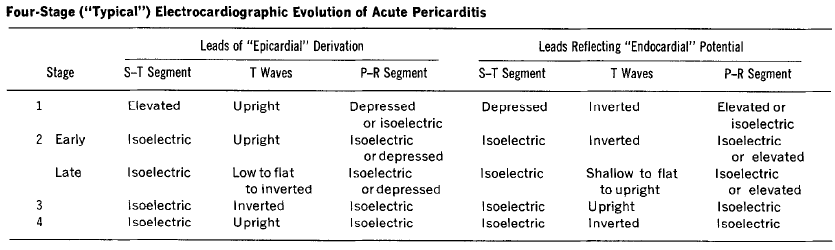
Spodick stages of pericarditis (1974)
Pericarditis is classically associated with ECG changes that evolve through four stages.
- Stage 1 – widespread STE and PR depression with reciprocal changes in aVR (occurs during the first two weeks)
- Stage 2 – normalisation of ST changes; generalised T wave flattening (1 to 3 weeks)
- Stage 3 – flattened T waves become inverted (3 to several weeks)
- Stage 4 – ECG returns to normal (several weeks onwards)
NB. Less than 50% of patients progress through all four classical stages and evolution of changes may not follow this typical pattern.
1974 – Spodick described the classic four-stage evolution of electrocardiographic changes with pericarditis, including ST elevations and PR depressions
Spodick sign in acute pericarditis (1974)
…the electrocardiographic registration of atrial injury in acute pericarditis is analogous to that of the stage 1 ventricular subepicardial injury in that each follows the orientation of its respective normal recovery (T) wave.
Since most atrial tissue lies posteriorly, to the right, and relatively cephalad of the ventricles, the orientation of a vector of generalized atrial injury (AP-R) would be expected to be to the right and superior (as shown by the frontal plane AP) and also posterior. The predominant P-R segment depressions across the precordium document the posterior component.
Because of these P-R segment deviations, it is of practical importance in acute pericarditis to consider the T-P segment to be the electrocardiographic base line to avoid mistaking P-R segment depression for S-T segment elevation.
Spodick 1974
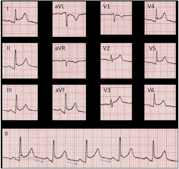
Clinical Case example: Uraemic pericarditis

Spodick sign in aortic stenosis (1976)
Auscultation of aortic stenosis murmur over the mid-clavicle
Clavicular transmission of aortic valve murmurs was investigated in 15 consecutive patients. In all patients, transmission to the clavicle of aortic systolic murmurs was detected both by auscultation and by phonocardiographic studies.
Clavicular auscultation regularly discloses aortic valve murmurs. The aortic systolic murmurs are consistently propagated to the clavicle, where they are usually conspicuously amplified. Aortic systolic murmurs are less frequently transmitted to the carotid and subclavian arteries, where they are usually considerably attenuated. Clavicular auscultation appears to be more rewarding than the traditional search for transmission of aortic murmurs to the carotid artery.
Spodick 1976
Major Publications
- Spodick DH. Acute Pericarditis Grune & Stratton. 1959.
- Spodick DH. Diagnostic electrocardiographic sequences in acute pericarditis. Significance of PR segment and PR vector changes. Circulation. 1973 Sep;48(3):575-80.
- Spodick DH. Electrocardiogram in acute pericarditis. Distributions of morphologic and axial changes by stages. Am J Cardiol. 1974 Apr;33(4):470-474. [Spodick sign]
- Spodick DH. Differential characteristics of the electrocardiogram in early repolarization and acute pericarditis. N Engl J Med. 1976 Sep 2;295(10):523-6.
- Spodick DH, Kerigan AT, de la Paz LR, Shahamatpour A, Kino M. Clavicular Auscultation. Preferential clavicular transmission and amplification of aortic valve murmurs. Chest. 1976 Sep;70(03):337-40. [Spodick sign, aortic stenosis]
- Spodick DH. Randomize the first patient: scientific, ethical, and behavioral bases. Am J Cardiol. 1983 Mar 1;51(5):916-7.
- Spodick DH. Comprehensive electrocardiographic analysis of acute myocardial infarction by individual and combined waveforms. Am J Cardiol. 1988 Sep 1;62(7):465-7.
- Spodick DH. Flow-directed pulmonary artery catheterization. Moratorium vs clinical trial. Chest. 1989 Mar;95(3):489-90.
- Spodick DH, Raju P, Bishop RL, Rifkin RD. Operational definition of normal sinus heart rate. Am J Cardiol. 1992 May 1;69(14):1245-6
- Spodick DH. The Pericardium: A Comprehensive Textbook. CRC Press. 1996
- Spodick DH. Acute cardiac tamponade. N Engl J Med. 2003 Aug 14;349(7):684-90.
- Mehrzad R, Spodick DH. Interatrial block: a virtual pandemic requiring attention. Iran J Med Sci. 2014 Mar;39(2):84-93
- Chhabra L, Spodick DH. Is It Time for a “Reverse Paradigm Shift” in the Treatment of Acute Idiopathic Pericarditis? Rev Esp Cardiol (Engl Ed). 2019 Sep;72(9):703-704.
References
Biography
- Saperia GM, Bishop RL. Profile in Cardiology. David H. Spodick. Clin Cardiol. 2004 Sep;27(9):533-4.
- Chhabra L. David H. Spodick, MD (1927 to 2019). Am J Cardiol 2019; 124(7): 1159-1160
- Chhabra L. Loss of a cardiology legend: A tribute to Professor David H. Spodick (1927-2019). J Electrocardiol. 2019 Sep-Oct;56:125-127.
- Bibliography. Spodick, David H. WorldCat Identities
Eponyms
- Chaubey VK, Chhabra L. Spodick’s Sign: A Helpful Electrocardiographic Clue to the Diagnosis of Acute Pericarditis. Perm J 2014; 18(1): e122
Eponym
the person behind the name
BA MA (Oxon) MBChB (Edin) FACEM FFSEM. Emergency physician, Sir Charles Gairdner Hospital. Passion for rugby; medical history; medical education; and asynchronous learning #FOAMed evangelist. Co-founder and CTO of Life in the Fast lane | On Call: Principles and Protocol 4e| Eponyms | Books |