ECG Case 062
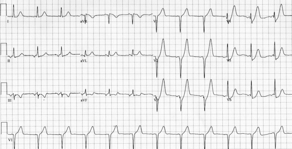
Middle aged patient presenting with chest pain and diaphoresis. Describe and interpret his ECG

Describe and interpret this ECG
ECG ANSWER and INTERPRETATION
Main Abnormalities
- ST depression in V2-6, which slopes upwards and joins the ascending limb of the T wave.
- Prominent, “rocket-shaped” T waves in the precordial leads V2-5.
- Subtle ST elevation in aVR.
Diagnosis
- This combination of ST depression with rocket-shaped T waves in the precordial leads V1-6 is referred to as the De Winter ECG pattern or “De Winter’s T waves” (also see ECG 019)
- It is becoming increasingly recognised as an anterior STEMI equivalent (~2% of LAD occlusions).
- Some authors are now recommending that this ECG pattern be treated identically to anterior STEMI, with urgent PCI or thrombolysis.

References
Further Reading
- Wiesbauer F, Kühn P. ECG Mastery: Yellow Belt online course. Understand ECG basics. Medmastery
- Wiesbauer F, Kühn P. ECG Mastery: Blue Belt online course: Become an ECG expert. Medmastery
- Kühn P, Houghton A. ECG Mastery: Black Belt Workshop. Advanced ECG interpretation. Medmastery
- Rawshani A. Clinical ECG Interpretation ECG Waves
- Smith SW. Dr Smith’s ECG blog.
- Wiesbauer F. Little Black Book of ECG Secrets. Medmastery PDF
TOP 100 ECG Series
Emergency Physician in Prehospital and Retrieval Medicine in Sydney, Australia. He has a passion for ECG interpretation and medical education | ECG Library |

Any comment about the rythm? The axis of the P wave (Negative in DII Positive in AVR) makes you think its an auricular rythm.
To me these resemble the Peaked T waves of Hyperkalemia?
Peaked T waves of hyperkalemia are narrow at but these are wide at the base then gradually narrows (rocket shaped).
I thought there were some pacing spikes especially lead 3