
![]()
ECG Case 061
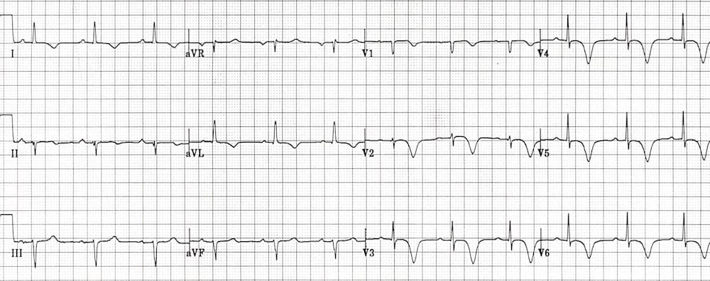
Middle-aged patient presenting with an episode of chest pain. Currently asymptomatic. Describe and interpret his ECG
Describe and interpret this ECG
ECG ANSWER and INTERPRETATION
This pattern of deeply inverted T waves in the anterolateral leads V2-6, I and aVL is characteristic of Wellens syndrome.
- This ECG pattern is highly predictive of a significant occlusive lesion of the LAD.
- The inverted T waves are a marker of reperfusion and may occur after an aborted anterior STEMI.
- Despite often being pain free and having normal cardiac enzymes at presentation, these patients are at risk of sudden LAD re-occlusion leading to massive anterior STEMI and are best managed with early angiography and PCI / CABG.
DIFFERENTIAL DIAGNOSIS
A similar pattern of deep anterolateral T-wave inversions may also be seen with:
- Apical hypertrophic cardiomyopathy – suspect if associated LVH
- Raised intracranial pressure – patient will be comatose (see Quiz ECG 012)
References
Further Reading
- Wiesbauer F, Kühn P. ECG Mastery: Yellow Belt online course. Understand ECG basics. Medmastery
- Wiesbauer F, Kühn P. ECG Mastery: Blue Belt online course: Become an ECG expert. Medmastery
- Kühn P, Houghton A. ECG Mastery: Black Belt Workshop. Advanced ECG interpretation. Medmastery
- Rawshani A. Clinical ECG Interpretation ECG Waves
- Smith SW. Dr Smith’s ECG blog.
- Wiesbauer F. Little Black Book of ECG Secrets. Medmastery PDF
TOP 100 ECG Series
Emergency Physician in Prehospital and Retrieval Medicine in Sydney, Australia. He has a passion for ECG interpretation and medical education | ECG Library |

This pattern is also seen in patient’s with takotsubu cardiomyopathy
We can notice inferior leads are negative and lateral leads are positive maybe left anterior fascicular block