
![]()
ECG Case 081
A 74 yr old patient presents to the ER having suffered several episodes of chest pain over the preceding 24 hours. His past medical history includes hypertension, hyperlipidaemia, diabetes, and ischaemic cardiac disease.
A series of ECG’s are performed on this patient.
ECG 1: taken on arrival to the Emergency Department with the patient pain free.

ECG 2: The patient then developed chest pain and the following ECG was recorded.
The episode of pain lasted only several minutes and resolved spontaneously.
ECG 3: taken 8 minutes after the second ECG with the patient now pain free.
Describe and interpret the ECGs
ECG ANSWER – ECG 1
This first ECG was taken on arrival to the Emergency Department with the patient pain free.
Rate:
- 66
Rhythm:
- Regular
- Sinus rhythm
Axis:
- Borderline LAD (~ -30 deg)
Intervals:
- PR – Normal (~180ms)
- QRS – Normal (80ms)
- QT – 400ms (QTc Bazett ~ 420 ms)
Segments:
- Minor ST depression lead III
Additional:
- T wave inversion leads II, III, aVF, V4, V5, V6
- Biphasic T wave leads aVR, V3
- Early precordial transition between V1 and V2
- Dominant R wave V2
- Prominent T wave lead V2
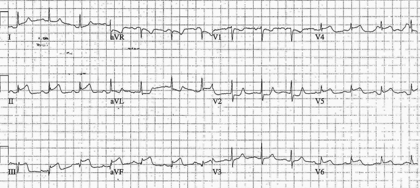
ECG ANSWER – ECG 2
The patient then developed chest pain and the following ECG was recorded.
Rate:
- 84
Rhythm:
- Regular
- Sinus rhythm
Axis:
- Normal (~20 deg)
Intervals:
- PR – Normal (~200ms)
- QRS – Normal (80ms)
- QT – 360ms (QTc Bazett ~ 415 ms)
Segments:
- ST Elevation leads II (2mm), III (3mm), aVF (2mm), V4 (1.5mm), V5 (1mm), V6 (1mm)
- ST Depression aVR, aVL, V1, V2
- Note horizontal ST morphology in V2
Additional:
- Pseudonormalisation of T waves leads II, III, aVF, V4, V5, V6
- Early precordial transition between V1 and V2
- Dominant R wave V2
- Prominent T wave lead V2
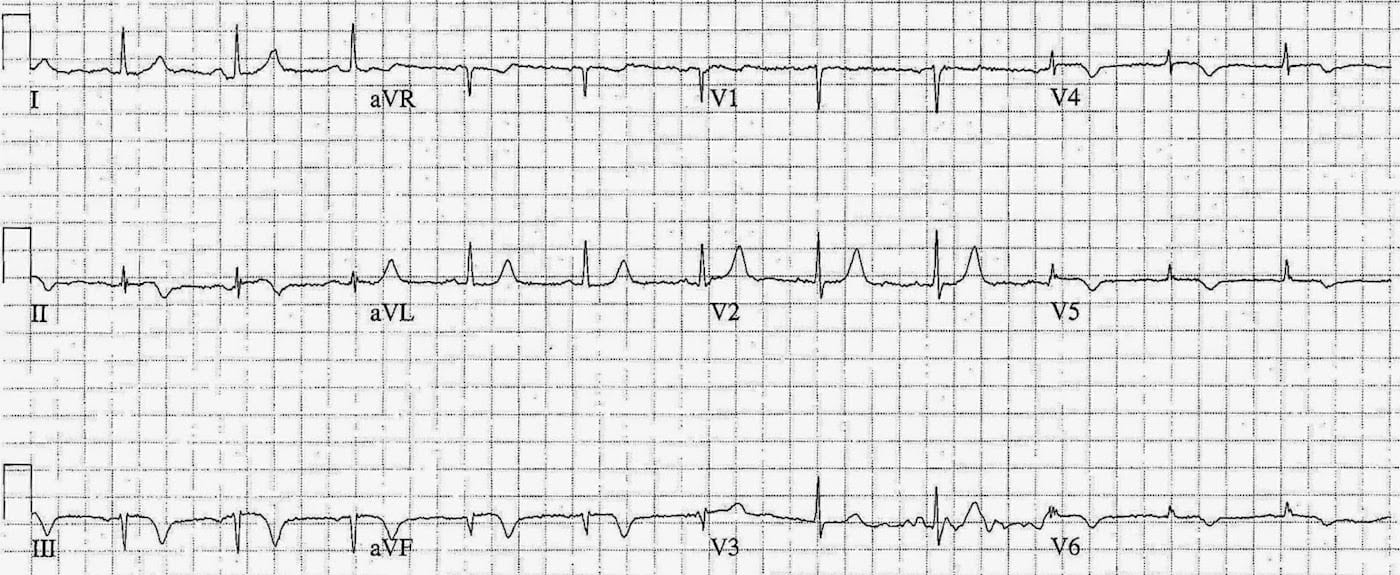
ECG ANSWER – ECG 3
The episode of pain lasted only several minutes and resolved spontaneously. This ECG was taken 8 minutes after the second ECG with the patient now pain free.
Rate:
- 66
Rhythm:
- Regular
- Sinus rhythm
Axis:
- Borderline LAD (~ -30 deg)
Intervals:
- PR – Normal (~180ms)
- QRS – Normal (80ms)
- QT – 380ms (QTc Bazett ~ 410 ms)
Segments:
- ST elevation leads III, aVF
- Reduced compered with ECG 2
Additional:
- T wave inversion leads II, III, aVF, V4, V5, V6
- Early precordial transition between V1 and V2
- Dominant R wave V2
- Prominent T wave lead V2
INTERPRETATION
ECG series showing ischaemia with re-perfusion (ECG 1), subsequent re-occlusion (ECG 2) with infero-postero-lateral STEMI, and spontaneous re-perfusion (ECG 3).
OUTCOME
The patient was immediately discussed with cardiology services. Treated with aspirin, clopidogrel, and placed on a heparin infusion and admitted to CCU. The patient remained pain free, troponin peaked at 12 hours, 4.8 (normal <0.05), and the patient was transfer the next day for angiography.
The angio showed:
- Right coronary: 98% stenosis –> stented
- Circumflex: 80% stenosis
- Left anterior descending: 80% proximal stenosis
- Left main: 20% proximal stenosis
- Left ventricle: Inferior hypokinesis with normal LV function
Check out the references belwo for some more great examples of re-perfusion / re-occlusion ECGs.
FURTHER READING
TOP 100 ECG Series
Emergency Medicine Specialist MBChB FRCEM FACEM. Medical Education, Cardiology and Web Based Resources | @jjlarkin78 | LinkedIn |
