


ECG Case 139
Dizzy
A 71-year-old man is brought to the emergency department after feeling persistently light-headed and unwell at home for the past hour.
He is alert and conversant, with no chest pain or breathlessness at rest. An ECG is recorded on arrival.
Questions
What is the most likely diagnosis?
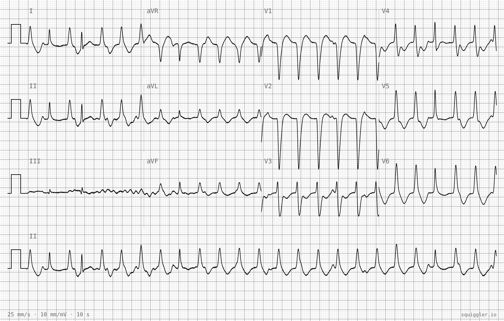
This is a regular broad-complex tachycardia at 143 beats per minute with a QRS duration of about 170 ms. In a 71-year-old man, a regular broad-complex tachycardia is ventricular tachycardia until proven otherwise. The rhythm strip contains capture and fusion beats, proving atrioventricular dissociation, which in a broad-complex tachycardia establishes VT. SVT with aberrancy can produce a broad QRS but cannot produce AV dissociation.
Which feature on this tracing confirms the diagnosis?
Capture beats are narrow complexes that appear when a sinus impulse conducts to the ventricles between tachycardia beats. A fusion beat has an intermediate morphology produced by simultaneous activation from both the sinus impulse and the ventricular focus. Together they prove that the atria and ventricles are operating independently, which in a broad-complex tachycardia effectively proves ventricular tachycardia. A wide QRS alone does not exclude aberrant conduction.
Which feature on this tracing confirms the diagnosis?
The 2022 ESC ventricular arrhythmia guideline recommends synchronised electrical cardioversion as first-line treatment for haemodynamically tolerated sustained monomorphic VT with known or suspected structural heart disease, provided the sedation risk is acceptable (Class I). Intravenous procainamide should be considered (Class IIa) and intravenous amiodarone may be considered (Class IIb). The reflex to reach for amiodarone because the patient is stable is common but is a weaker recommendation than cardioversion. The Resuscitation Council UK adult tachycardia algorithm still names amiodarone for stable broad-complex tachycardia, so practice varies between guidelines.
Follow up and review
Synchronised DC cardioversion is performed under procedural sedation in the resuscitation room and sinus rhythm is restored. A repeat ECG is recorded.
Questions
What does this post-cardioversion ECG show?
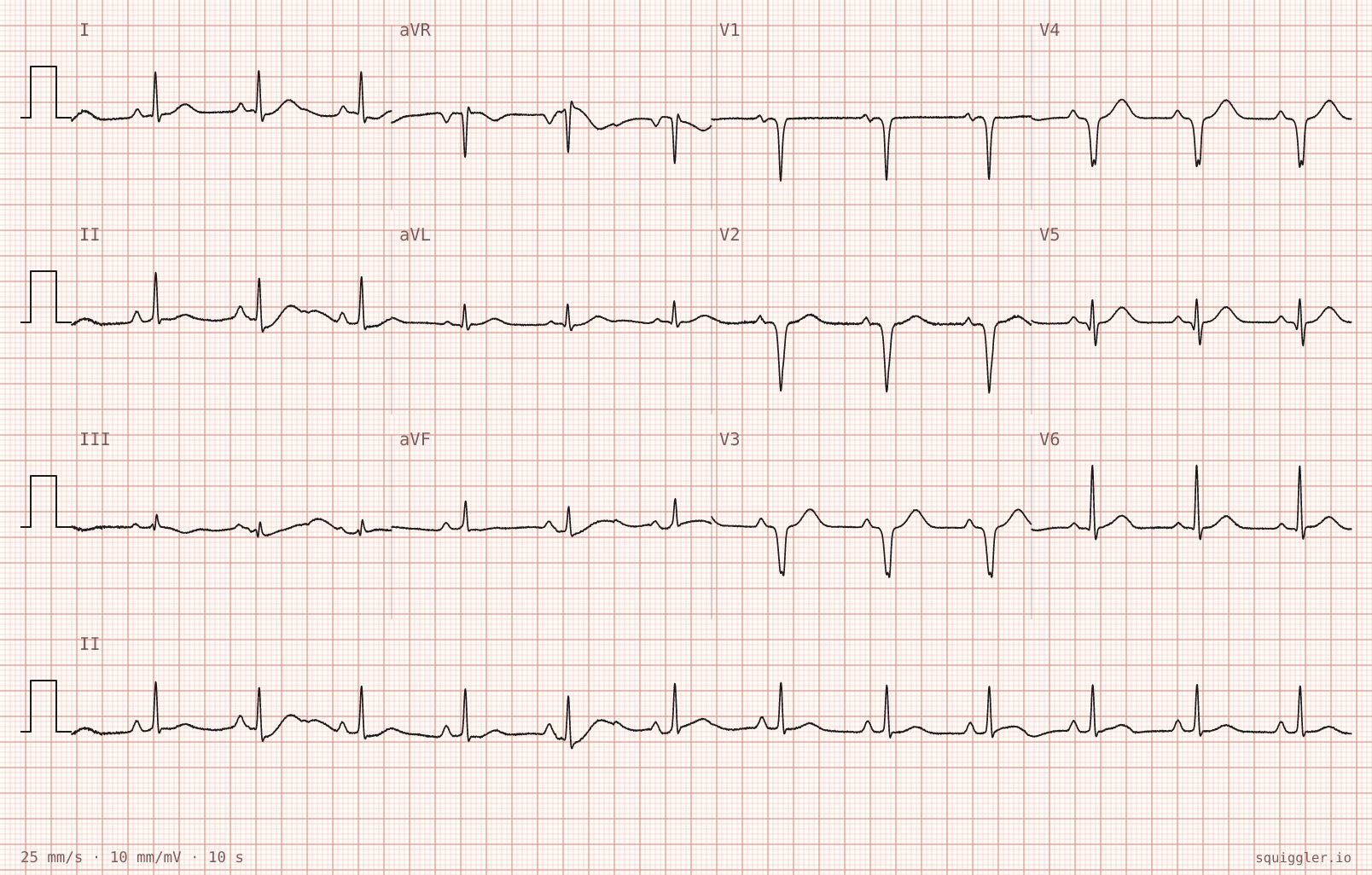
There are QS complexes across V1 to V4 with no R-wave progression through the anterior chest leads, indicating a previous anterior myocardial infarction. The T waves are upright in V3 to V6 and there is no acute ST elevation, distinguishing this from an acute event. The QRS is narrow at 74 ms, excluding a bundle branch block.
Why is this finding important in this patient?
Monomorphic ventricular tachycardia in the setting of a healed myocardial infarction is typically scar-mediated re-entry. The infarct scar provides the slow conduction zone that sustains the tachycardia circuit. This patient requires cardiology assessment, echocardiography to assess left ventricular function, and consideration of an implantable cardioverter-defibrillator for secondary prevention of sustained VT.
Diagnosis and Summary
Monomorphic ventricular tachycardia with old anterior myocardial infarction
The presence of capture and fusion beats in the rhythm strip proves atrioventricular dissociation and confirms ventricular tachycardia. Haemodynamic stability does not distinguish VT from supraventricular tachycardia with aberrant conduction, and treating a stable broad-complex tachycardia as SVT is a well-recognised and dangerous error.
For haemodynamically tolerated monomorphic VT with suspected structural heart disease, the 2022 ESC guideline recommends synchronised electrical cardioversion as first-line treatment. After cardioversion, the resting ECG reveals QS complexes across the anterior chest leads, indicating a previous anterior myocardial infarction. The infarct scar is the substrate for re-entrant VT. This patient needs cardiology assessment, echocardiography and consideration of an implantable cardioverter-defibrillator for secondary prevention.
References
- Brugada P, Brugada J, Mont L, Smeets J, Andries EW. A new approach to the differential diagnosis of a regular tachycardia with a wide QRS complex. Circulation. 1991 May;83(5):1649-59
- Wellens HJ. Electrophysiology: Ventricular tachycardia: diagnosis of broad QRS complex tachycardia. Heart. 2001 Nov;86(5):579-85.
- Zeppenfeld K, Tfelt-Hansen J, de Riva M, et al. 2022 ESC Guidelines for the management of patients with ventricular arrhythmias and the prevention of sudden cardiac death. Eur Heart J. 2022;43(40):3997-4126.
Note. These ECG tracings were generated, not recorded. The Squiggler ECG generation studio can be used to live edit and review each generated ECG for education purposes. Open the studio
TOP 150 ECG Series

David Yu, M.D., Ph.D. Full time anesthesiologist in the department of Perioperative medicine and Intensive Care (PMI), Karolinska University Hospital, Sweden. Dual board certified in internal medicine, and anesthesiology and critical care. Clinical interests are the critically ill patient, hemodynamic cases, perioperative echocardiography and procedural awareness, and of course all things ECG! | Squiggler |