


Freiberg infraction
Freiberg infraction (Freiberg disease; Köhler disease II; Freiberg–Köhler disease) is an osteochondrosis of a lesser metatarsal head, characterised by subchondral osteonecrosis, collapse and progressive deformity of the articular surface. The second metatarsal is most commonly affected, followed by the third; fourth-metatarsal involvement is uncommon.
Freiberg infraction classically presents during adolescence, particularly in females, although both sexes and older patients may be affected. Trnka reports involvement of the second metatarsal in approximately 68%, the third in 27%, and the fourth in 3% of cases.
Clinical presentation
Patients typically develop forefoot pain localised to the affected metatarsal head, worsened by weight bearing, walking barefoot, high heels or flexible-soled footwear. Examination may demonstrate focal tenderness, fusiform swelling, reduced metatarsophalangeal movement, dorsal impingement and occasionally crepitus.
Plain radiographs may be normal early in the disease. With progression there is sclerosis, flattening and fragmentation of the metatarsal head, widening followed later by narrowing of the metatarsophalangeal joint, osteochondral loose bodies and secondary arthrosis. MRI can demonstrate early marrow abnormality and osteonecrosis before established radiographic collapse.
Pathology and aetiology
The precise aetiology remains uncertain and probably multifactorial. Proposed contributors include repetitive mechanical loading or trauma, vascular compromise, altered forefoot biomechanics, skeletal maturation and genetic susceptibility. Vascular injection studies demonstrate a potentially vulnerable extraosseous arterial network around the lesser metatarsal heads.
The dorsal portion of the metatarsal head is most affected. Subchondral bone failure and osteonecrosis lead to collapse of the dorsal articular surface, while the plantar articular cartilage is characteristically preserved until later stages. Progressive collapse may produce fragmentation, loose bodies and ultimately degenerative metatarsophalangeal arthrosis.
Management
Initial treatment is non-operative, aimed at unloading the affected metatarsal and reducing metatarsophalangeal stress. Options include activity modification, analgesia, metatarsal off-loading or orthoses, and stiff-soled or rocker-bottom footwear. Early lesions may heal and remodel spontaneously.
Surgery is reserved for persistent symptoms despite conservative treatment or progressive structural disease. Earlier-stage procedures aim to preserve the joint and viable plantar cartilage and include core decompression, debridement and metatarsal osteotomy, particularly dorsal closing-wedge or modified Weil osteotomy. Osteochondral grafting may be considered for selected focal defects.
Advanced disease with irreversible collapse and arthrosis may require resection or interposition arthroplasty, although modern practice generally favours joint-preserving procedures where possible.
History of the Freiberg infraction
1903 – Albert Henry Freiberg (1868–1940) encountered the first of a series of patients with pain localised to the second metatarsophalangeal joint and characteristic deformity of the second metatarsal head. His first patient was a 16-year-old girl who attributed the onset to “stubbing” her foot while playing tennis.
1913 – Freiberg presented six cases to the Southern Surgical and Gynecological Association. He believed the appearances represented an infraction of the distal second metatarsal, regarding trauma or repeated mechanical injury as the likely cause.
During the past ten years I have encountered six cases of infraction of the distal end of the second metatarsal bone. The patient was a girl aged sixteen years. She had been suffering from pain in the ball of the foot for about six months. The patient was quite sure that the condition dated from and was due to a game of tennis in which she “stubbed her foot“
Freiberg 1913
1914 – Freiberg published Infraction of the Second Metatarsal Bone: A Typical Injury. Radiographs demonstrated flattening and collapse of the metatarsal head, sometimes with loose osteochondral bodies and subsequent degenerative change. Four patients were managed conservatively and two underwent arthrotomy with removal of loose bodies.
I felt justified in the diagnosis of infraction of the distal end of the second metatarsal, a condition which I have thus far failed to find described in the literature.
Freiberg 1914
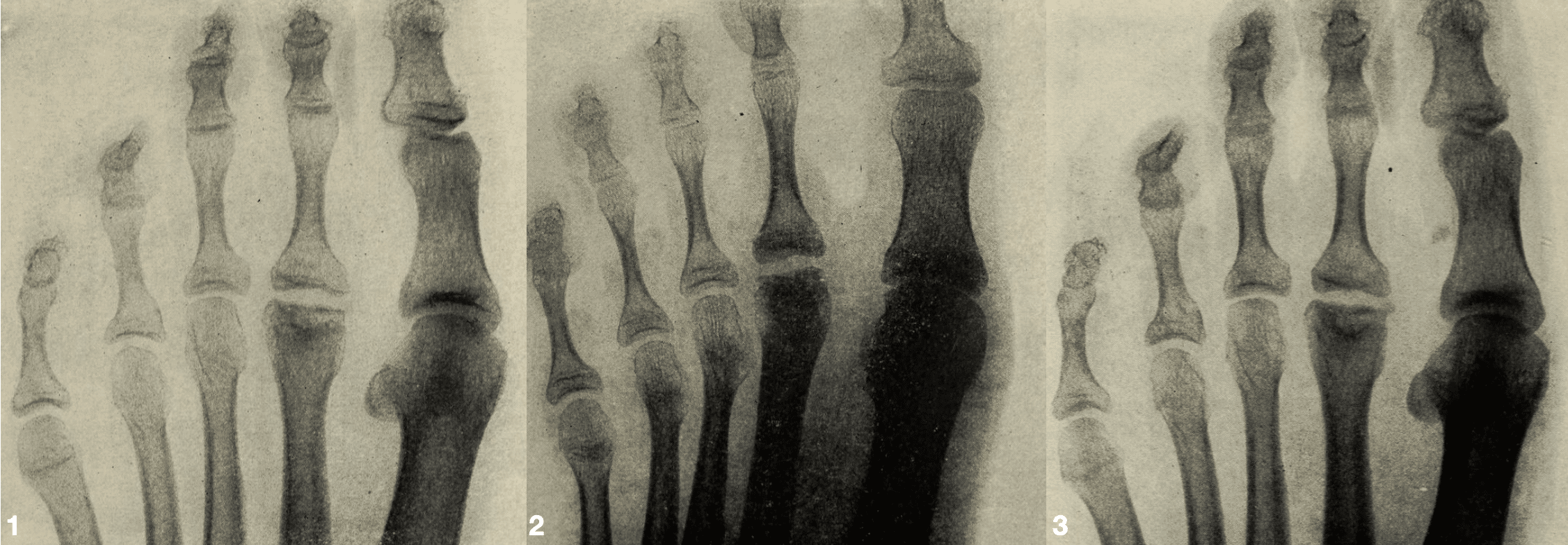
1915 – Alban Köhler (1874–1947), apparently unaware of Freiberg’s American report, included a brief description in the second edition of Grenzen des Normalen und Anfänge des Pathologischen im Röntgenbilde. He described a “most peculiar disease” of the second metatarsophalangeal joint characterised by flattening or depression of the articular surface, an apparent 1–2 mm fracture- or infraction-like cleft, and broadening of the distal metatarsal.
His wording that the disease had, “to the knowledge of the author,” not previously been represented in the literature makes an independent observation quite plausible rather than a deliberate claim against Freiberg.
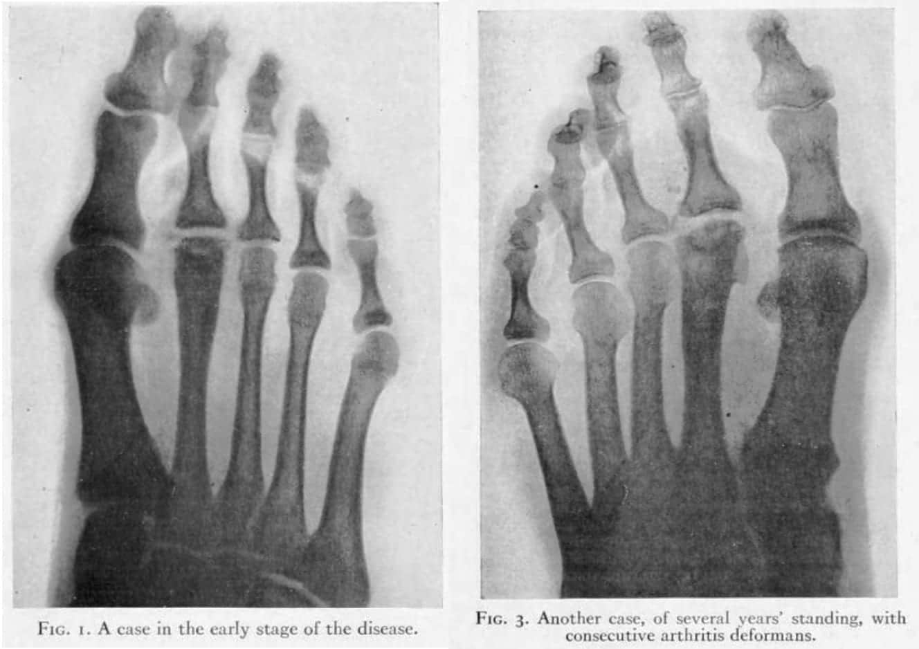
1917 – Willis C. Campbell published Infraction of the Head of the Second and Third Metatarsal Bones. He credited Freiberg with first defining the condition in 1914 and favoured a mechanical-traumatic explanation. Campbell reported involvement of the third metatarsal head, demonstrating that the disorder was not confined to the second metatarsal. One patient had symptomatic third-metatarsal disease in one foot and previous second-metatarsal involvement in the opposite foot.
1920 – Köhler presented a much more detailed analysis Eine typische Erkrankung des 2. Metatarsophalangealgelenkes to the 11th congress of the German Roentgen Ray Society. This was published in the Münchener medizinische Wochenschrift in 1920 with the English translation appeared in the American Journal of Roentgenology in 1923.
Köhler expanded the radiographic picture beyond collapse of the metatarsal head. He emphasised irregular widening of the joint space, shortening and flattening of the head, thickening of the distal metatarsal and progressive obliteration of the neck. He also challenged a simple traumatic origin:
The joint-space is not only broadened, but the broadening is strikingly irregular, so that the fibular half of the space is often double that of the tibial half (…) The head of the metatarsal is undoubtedly shortened, not in toto but in its distal third, as if the cap had been driven in.
About 1 patient in 10 gave a history of trauma. We need not dwell further on this point. Certainly trauma alone cannot be the cause.
Köhler, 1920
1923 – Köhler’s 1920 account appeared in English in the American Journal of Roentgenology as Typical disease of the second metatarsophalangeal joint. Philip Lewin published Juvenile deforming metatarsophalangeal osteochondritis. Freiberg’s infraction of the metatarsal head. Reviewing 65 reported cases, Lewin stated:
This condition is erroneously known as Köhler’s metatarsophalangeal syndrome.
Lewin credited Freiberg with the first description and proposed trauma, circulation and infection as possible contributing factors. He preferred the descriptive term juvenile deforming metatarsophalangeal osteochondritis
1924 – Köhler responded directly in Ueber die ersten Veröffentlichungen der typischen Erkrankung des zweiten Metatarsophalangealgelenks. Now aware of Freiberg’s paper through Lewin and the editorial note appended to the English translation of his work, Köhler conceded the publication priority:
Freiberg’s paper was therefore printed several months earlier.
He went further, stating that because Freiberg had recognised and described the lesion independently, Freiberg’s name should henceforth be mentioned first.
However, Köhler argued that Freiberg had described primarily the infraction or fracture component, whereas his own later work defined a broader radiographic syndrome. He emphasised the progressive thickening of the distal metatarsal with disappearance of the neck, which was visible even in Freiberg’s three published radiographs but had not been recognised by Freiberg.
1926 – Freiberg revisited the condition in The so-called infraction of the second metatarsal bone. With the benefit of subsequent cases and Köhler’s observations, he acknowledged that simple trauma was no longer a satisfactory explanation of the clinical and radiographic syndrome.
He then stepped away from the priority dispute (see Eponymythology below)
The matters of priority of publication and of the attachment of an eponymic title to the condition seem to me of little importance.
1928 – In Röntgenology: The Borderlands of the Normal and Early Pathological in the Skiagram, Köhler reviewed the accumulated experience of more than 100 reported cases. He regarded the characteristic disease pattern as extending beyond metatarsal-head collapse to include joint-space widening and irregularity, flattening of the articular cap, capsular calcification, shortening of the head, and particularly progressive thickening of the distal metatarsal with obliteration of the neck.
Köhler reiterated that the disorder could not be adequately understood as a simple traumatic “infraction”, while distinguishing these characteristic changes from the later development of secondary arthritis deformans.
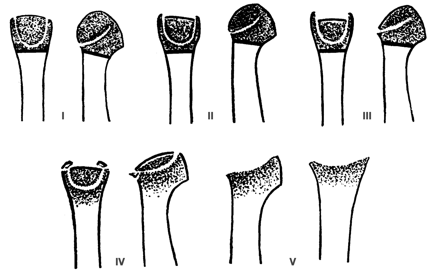
1967 – Ian Scott Smillie (1907-1992) described the five-stage classification that remains the most widely recognised system for Freiberg disease. His stages were based on operative observations of progressive structural failure of the metatarsal head.
It is possible by timely surgical intervention to interrupt the pathological process of Freiberg’s infraction and restore the conformation of the metatarsal head. For the purposes of treatment, the natural history of the disease can be conveniently divided into five stages
Smillie 1967
1979 – Gauthier and Elbaz reframed Freiberg disease as a subchondral bone fatigue fracture, with subsequent loss of vascular continuity, osteonecrosis and collapse. They also introduced a dorsal closing-wedge osteotomy designed to rotate preserved plantar cartilage into the principal weight-bearing surface. Their series included 88 affected metatarsals, with 53 treated by the new osteotomy.
1987–2005 – Subsequent studies expanded the concept beyond plain radiography. Scintigraphy demonstrated early patterns compatible with osteonecrosis, and MRI allowed identification before established metatarsal-head collapse. Vascular injection studies demonstrated the potentially vulnerable arterial network of the lesser metatarsal heads and occurrence in identical twins raised the possibility of genetic predisposition.
These observations contribute to the current multifactorial model incorporating biomechanical overload, vascular compromise, skeletal maturation and individual susceptibility.
Eponymythology
Who described it first?
Freiberg has priority of publication. He presented his series in 1913 and published six cases in 1914. Köhler prepared his initial description independently in 1913–1914, but it did not appear until the second edition of Grenzen des Normalen in 1915. When the chronology was brought to his attention, Köhler acknowledged that Freiberg’s paper had appeared several months earlier and stated that Freiberg’s name should be mentioned first.
Why Köhler disease II?
The disagreement was less about who published first than about what constituted the disease. Freiberg’s original concept centred on traumatic infraction of the metatarsal head. Köhler argued that this represented only part of a broader radiographic syndrome and particularly emphasised distal metatarsal thickening with obliteration of the neck, together with abnormalities of the joint space and adjacent articular surfaces. In 1923, Philip Lewin challenged the existing attribution directly, writing that the condition was:
…erroneously known as Köhler’s metatarsophalangeal syndrome.
Köhler accepted Lewin’s point about publication priority, but rejected the implication that his contribution was merely a later duplication of Freiberg’s. He maintained that his 1920 work had established the more complete radiographic phenotype.
Whose name survived?
English-language usage favours Freiberg disease or Freiberg infraction, while Köhler disease II, Freiberg–Köhler disease and Köhler–Freiberg disease persist as alternative terms. Freiberg showed little enthusiasm for the contest, writing in 1926:
The matters of priority of publication and of the attachment of an eponymic title to the condition seem to me of little importance.
The eponym therefore records two different contributions rather than a genuine priority tie: Freiberg supplied the first published description whilst Köhler independently recognised the lesion and subsequently broadened its radiographic definition.
Infraction, not infarction
Freiberg deliberately used infraction, an older term for an incomplete or non-displaced fracture, reflecting his original traumatic interpretation. Modern concepts emphasise osteochondrosis, osteonecrosis and multifactorial pathogenesis, but the historical term Freiberg infraction remains in use.
Associated Persons
- Albert Henry Freiberg (1868-1940)
- Alban Köhler (1874–1947)
- Ian Scott Smillie (1907-1992)
Alternative names
- Freiberg disease, Freiberg infraction, Freiberg’s infraction
- Köhler disease II
- Freiberg-Köhler syndrome
References
Original articles
- Freiberg AH. Infraction of the second metatarsal – a typical injury. Surgery, gynecology & obstetrics 1914; 19(2): 191-193
- Freiberg AH. Infraction of the second metatarsal – a typical injury. Transactions of the Southern Surgical and Gynecological Association 1914; 26: 171-174.
- Köhler A. Grenzen des Normalen und Anfänge des Pathologischen im Röntgenbilde. 1915
- Campbell WC. Infraction of the Head of the Second and Third Metatarsal Bones: Report of Cases, American journal of orthopedic surgery. 1917; 15: 721
- Lewin P. Juvenile deforming metatarsophalangeal osteochondritis. Freiberg’s infraction of the metatarsal head. JAMA 1923;81;(3):189-192.
- Köhler A. Eine typische Erkrankung des 2. Metatarsophalangealgelenkes. Munchener medizinische Wochenschrift, 1920; 67: 1289-1290 [Typical disease of the second metatarsophalangeal joint. Translation: American journal of roentgenology. 1923, 10: 705-710]
- Köhler A. Ueber die ersten Veröffentlichungen der typischen Erkrankung des zweiten Metatarsophalangealgelenks. Münchener medizinische Wochenschrift, 1924; LXXI(4): 109
- Freiberg AH. The so-called infraction of the second metatarsal bone. J Bone Joint Surg 1926; 8: 257-61.
- Köhler A. Röntgenology: The Borderlands of the Normal and Early Pathological in the Skiagram. Trans. Arthur Turnbull. William Wood. 1928
Review articles
- Smillie IS. Treatment of Freiberg’s infraction. Proc R Soc Med. 1967 Jan;60(1):29-31
- Gauthier G, Elbaz R. Freiberg’s infraction: a subchondral bone fatigue fracture. A new surgical treatment. Clin Orthop Relat Res. 1979 Jul-Aug;(142):93-5
- Binek R, Levinsohn EM, Bersani F, Rubenstein H. Freiberg disease complicating unrelated trauma. Orthopedics. 1988; 11(5): 753-7.
- Scartozzi G, Schram A, Janigian J. Freiberg’s infraction of the second metatarsal head with formation of multiple loose bodies. J Foot Surg. 1989; 28(3): 195-9.
- Katcherian DA. Treatment of Freiberg’s disease. Orthop Clin North Am. 1994; 25(1): 69-81
- Ashman CJ et al. Forefoot Pain Involving the Metatarsal Region: Differential Diagnosis with MR Imaging. Radiographics. 2001; 21(6): 1425-1440
- Torriani M, Thomas BJ, Bredella MA, Ouellette H. MRI of metatarsal head subchondral fractures in patients with forefoot pain. AJR Am J Roentgenol. 2008; 190(3): 570-5.
- Cerrato RA. Freiberg’s disease. Foot Ankle Clin. 2011; 16(4): 647-58.
- Lin HT, Liu AL. Freiberg’s infraction. BMJ Case Rep. 2013 Jun 18;2013:bcr2013010121
- Talusan PG, Diaz-Collado PJ, Reach JS Jr. Freiberg’s infraction: diagnosis and treatment. Foot Ankle Spec. 2014 Feb;7(1):52-6
- Trnka HJ, Lara JS. Freiberg’s Infraction: Surgical Options. Foot Ankle Clin. 2019 Dec;24(4):669-676
- Inokuchi R, Iwashita K, Jujo Y, Takao M. Freiberg’s disease. BMJ Case Rep. 2019 Oct 10;12(10):e232171
- Cadogan M. Eponymythology of foot injuries. LITFL
eponymictionary
the names behind the name

MB BAO BCh Trinity College Dublin, MSc Clinical Data Analytics University of Galway. Junior doctor in Perth, doctoral researcher in MRI radiomics. Working towards radiology training.

BA MA (Oxon) MBChB (Edin) FACEM FFSEM. Emergency physician, Sir Charles Gairdner Hospital. Passion for rugby, medical history, medical education, and asynchronous learning #FOAMed evangelist. Editor-in-Chief and Webmaster of Life in the Fast lane, LITFL | On Call 4e| Eponyms | Books | Horology |