


Alban Köhler
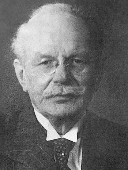
Alban Köhler (1874-1947) was a German Radiologist.
Köhler was a pioneer of osteoradiology. He was among the first physicians to devote his career principally to radiology and became particularly influential in the radiographic interpretation of bones and joints.
Köhler combined meticulous anatomical investigation with clinical radiography. His studies of the hip established the Köhler teardrop and the pelvic-wall contour the Köhler line. He also described the childhood navicular osteochondrosis now known as Köhler disease, contributed to the description of Freiberg–Köhler disease, and documented the medial femoral-condylar ossification subsequently called the Pellegrini-Stieda lesion.
His most influential publication, Grenzen des Normalen und Anfänge des Pathologischen im Röntgenbilde, first appeared in 1910. Conceived as a guide to distinguish normal variation from early disease, it became an international radiological reference work and continued through numerous German and English editions long after his death.
Köhler developed techniques in teleradiography, dosimetry and radiation protection and in 1909 introduced Gittertherapie (grid therapy), an early form of spatially fractionated radiotherapy.
Biography
- Born on March 1, 1874 in Petsa, Thuringia, into a farming family.
- 1893 – Completed secondary education (Abitur). Studied medicine at Freiburg, Leipzig, Erlangen and Berlin.
- 1897 – Awarded medical doctorate at Freiburg, following studies in Freiburg, Leipzig, Berlin and Erlangen. Dissertation on pituitary tumours.
- 1898 – Passed the German state medical examination. Worked as assistant to pathologist Christian Georg Schmorl (1861–1932) in Dresden, followed by military service in Freiburg and Dresden attaining the rank of Stabsarzt der Reserve.
- 1899–1902 – Assistant to surgeon Friedrich Cramer (1847–1903) at St Josef Hospital, Wiesbaden, specialising in the new field of radiology.
- 1901 – Published Knockenerkrankungen im Röntgenbilde, an atlas of bone disease on radiographs.
- 1902 – Established radiological practice in Paulinenstraße, Wiesbaden as the first German Facharzt für Röntgenologie (specialist in radiology).
- 1905 – Co-founded the Deutsche Röntgengesellschaft
- 1908 – Published description of childhood navicular osteochondrosis (Köhler disease I)
- 1909 – Introduced Gittertherapie (grid therapy), spatially fractionated X-ray treatment designed to deliver large doses to deep lesions while limiting skin injury.
- 1910 – Published Grenzen des Normalen und Anfänge des Pathologischen im Röntgenbilde
- 1912 – Elected first chairman (erster Vorsitzender) of the Deutsche Röntgengesellschaft and presided over the 8th German Roentgen Congress in Berlin.
- 1913 – Awarded the title Professor by Duke Ernst of Saxe-Altenburg, in recognition of his scientific work, through the medical faculty of the University of Jena.
- 1914–1918 – Served throughout the First World War as a radiologist in the reserve military hospitals of Wiesbaden and at the Großes Hauptquartier.
- 1920 – Published his account of disease of the second metatarsophalangeal, Köhler disease II.
- 1931 – At the radiology congress in Baden-Baden, urged colleagues with 20+ years of X-ray exposure to undergo blood-count surveillance
- 1933 – Awarded the Hermann Rieder Medal by the German Radiological Society at its 24th Congress in Bremen.
- 1934 – At the 4th International Congress of Radiology in Zurich, presented findings from an international survey of health problems among radiologists with prolonged occupational exposure.
- 1944 – Received the Goethe Medal. On his 70th birthday he reflected that he had spent 45 years “within the realm and spell of Röntgen’s wonder rays”.
- 1946 – Completed his memoirs Röntgenarztes Erdenwallen.
- Died suddenly of heart failure on February 26, 1947 in Nieder-Selters im Taunus, aged 72
Medical Eponyms
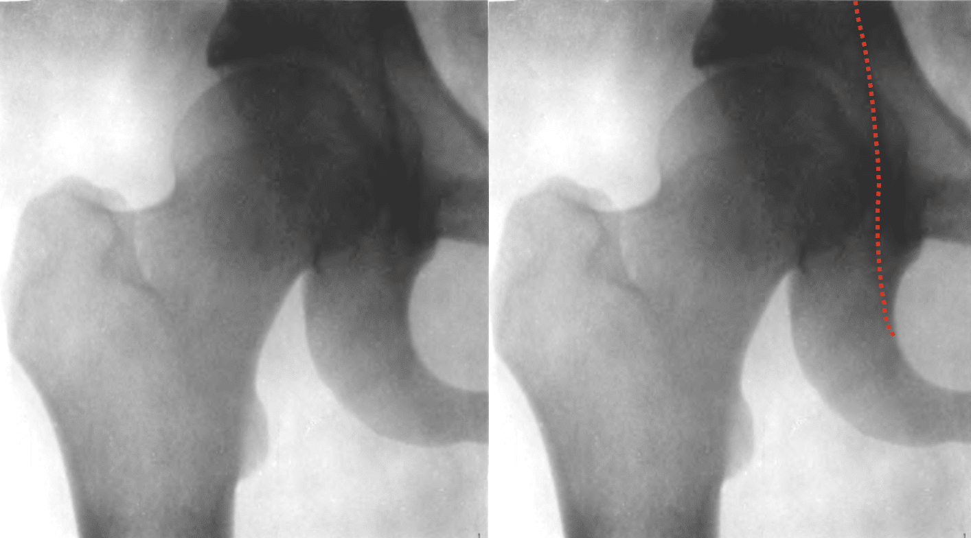
Köhler line (1905)
Köhler line (ilioischial line) is a radiographic landmark on the AP pelvis used to assess the medial relationship of the acetabulum and femoral head to the pelvis, particularly in evaluation of acetabular depth and protrusio acetabuli.
1905 – Köhler published Die normale und pathologische Anatomie des Hüftgelenks und Oberschenkels in röntgenographischer Darstellung. He investigated the projectional anatomy of the acetabulum and medial pelvic wall and identified the long pelvic-wall contour the Köhler line.
1910 – He formalised the projectional anatomy in Grenzen des Normalen, stating that the apparent lines represented tangential projections of curved pelvic surfaces rather than discrete anatomical edges.
Köhler teardrop (1905)
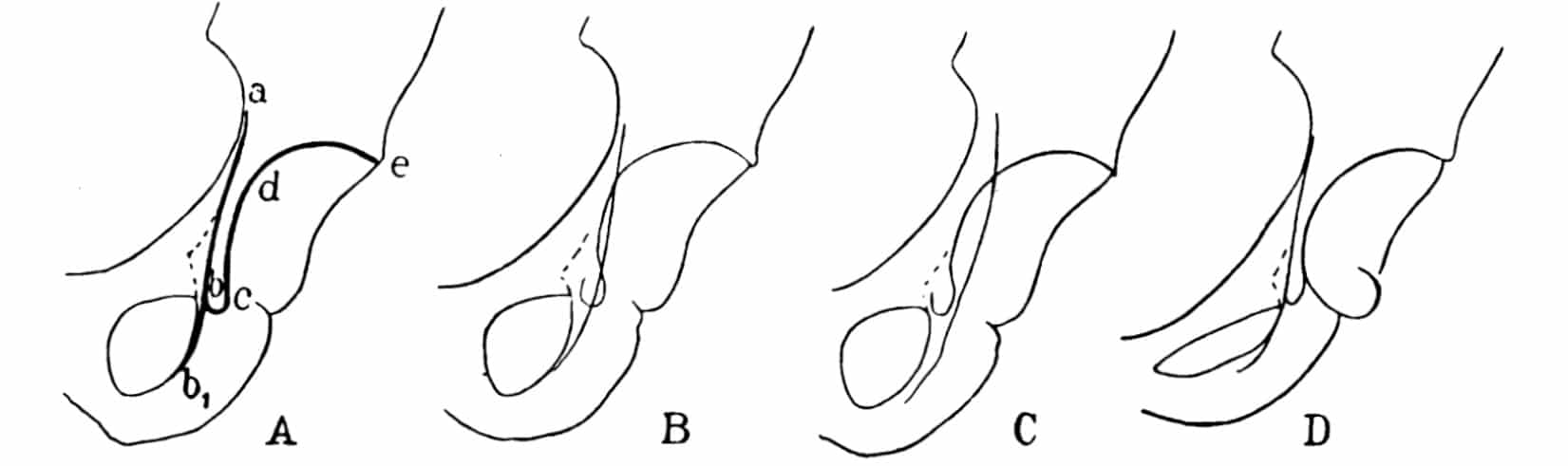
Köhler teardrop (Köhlersche Tränenfigur; pelvic teardrop; U-figure) is the U- or teardrop-shaped radiographic summation shadow medial to the acetabulum on an AP pelvis radiograph.
1905 – Köhler described the appearance of the teardrop and investigated how its form changed with pelvic position and X-ray projection. He compared it with a falling water drop or glass tear. Subsequent studies confirmed that the teardrop is a composite projection produced by several acetabular and pelvic cortical surfaces.
Köhler disease I (1908)
Köhler disease (Köhler disease I) is a self-limiting childhood osteochondrosis of the developing tarsal navicular, typically presenting with dorsomedial foot pain and limp.
1908 – Köhler described the condition in three boys aged 5–9 years in Uber eine häufige, bisher anscheinend unbekannte Erkrankung einzelner kindlicher Knochen. He reported on characteristic radiographic changes and their subsequent restoration. He described abnormalities of the navicular’s size, shape, architecture and calcification and emphasising the favourable prognosis.

Köhler-Pellegrini-Stieda lesion (Pellegrini-Stieda disease)
Pellegrini–Stieda lesion refers to post-traumatic ossification adjacent to the medial femoral condyle, usually involving the region of the medial collateral ligament.
Köhler illustrated such a lesion in his 1905 hip and femur monograph in a 56-year-old man with persistent knee pain after trauma. Augusto Pellegrini (1877–1958) described a similar lesion in the same year, and Alfred Stieda (1869–1945) subsequently provided a more detailed account, leading to the better-known Pellegrini–Stieda terminology.

Freiberg-Köhler disease [Köhler disease II] (1915, 1920)
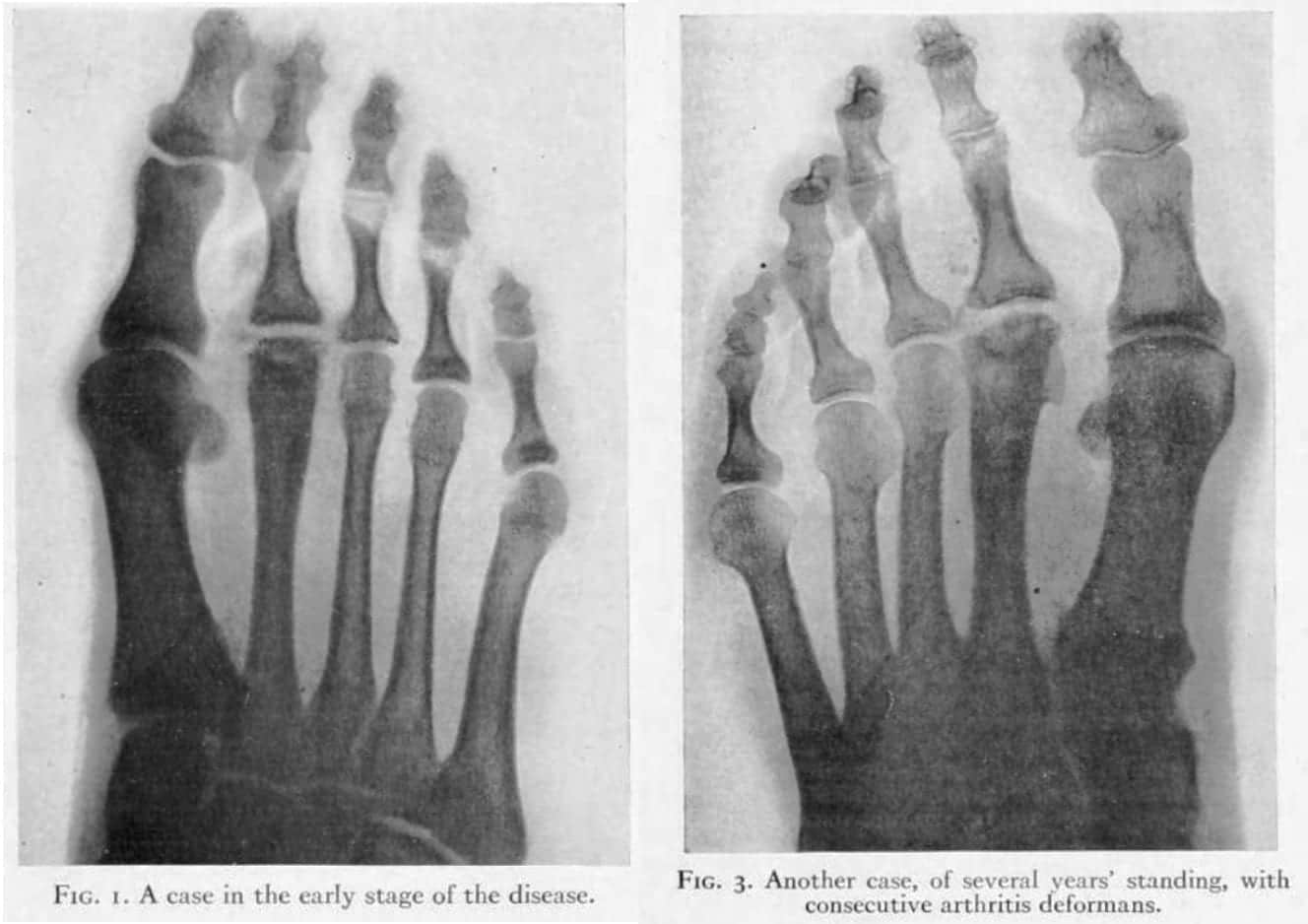
Freiberg–Köhler disease (Köhler disease II; Freiberg infraction) is an osteochondrosis of a lesser metatarsal head, most commonly the second, producing progressive flattening and collapse of the articular surface.
Albert Henry Freiberg (1868–1940) has publication priority, presenting cases in 1913 and publishing them in 1914. Köhler independently described the radiographic entity in the 1915 second edition of Grenzen des Normalen. Köhler expanded the syndrome in 1920 with Eine typische Erkrankung des 2. Metatarsophalangealgelenkes. He described abnormalities beyond the fracture-like lesion, including widening and irregularity of the joint space and progressive thickening of the distal metatarsal with loss of the normal neck contour.
Original
English
Am 2. Metatarso phalangeal-Gelenk kommt eine äusserst eigenartige Erkrankung vor, die, soviel Verfasser weiss, in ihrer Eigenart in der Literatur bisher nirgends gewürdigt worden ist. Es handelt sich um eine Abflachung oder Abplattung der Gelenkfläche des Köpfchens des betreffenden Metatarsus, die manchmal gelockert bez. etwas abgehoben zu sein scheint; jedenfalls sieht man mitunter, bei den anscheinend frischeren Fällen, einen infraktions- oder frakturartigen 1-2mm breiten, durchlässigen Zwischenraum (…)
Äusserst merkwürdig ist, dass die ganze distale Hälfte des befallenen Metatarsus distalwärts gleichmässig an Umfang zunimmt.
At the 2nd metatarso-phalangeal joint, a most peculiar disease may arise, which, to the knowledge of the author, has not been honoured in literature so far. It is a flattening, or squashing of the articular surface of the head of the affected metatarsal, which is sometimes loosened, or apparently somewhat lifted; in any case one may see in the fresher cases, an infraction- or fracture-like intermediary space of 1-2mm (…)
Key medical contributions
Pioneer of musculoskeletal radiology
Köhler was among the earliest physicians to make radiology of bones and joints a distinct area of expertise. His 1901 atlas Knockenerkrankungen im Röntgenbilde and 1905 monograph on the hip and femur were early attempts to correlate radiographic appearances with normal and pathological skeletal anatomy.
Borderlands of the Normal and Early Pathological
Köhler’s most influential work was Lexikon der Grenzen des normalen und der Anfänge des pathologischen im Röntgenbilde, first published in 1910. Köhler recognised that one of the greatest hazards in early radiology was mistaking normal variation for disease, or subtle early disease for normal anatomy. The book became an international reference work and was repeatedly revised and translated after his death.
Radiographic technique and cardiac imaging
Köhler contributed to the development of teleradiography, promoted improved techniques for radiographic measurement of the heart, introduced a double-plate method, and investigated stereoscopic and serial radiography. He devised practical methods for localisation of foreign bodies and was interested in radiographic demonstration of structures such as aortic calcification.
Grid therapy and radiotherapy
In 1909, Köhler introduced Gittertherapie (grid or sieve therapy), an early form of spatially fractionated radiotherapy. By shielding small regions of skin while irradiating intervening areas, he attempted to deliver high doses to deeper tumours while permitting the skin to recover. Often regarded as the first radiotherapist to develop and clinically test this principle.
Radiation dosimetry and protection
Köhler studied radiation injury from the early years of his career and introduced practical methods of dose estimation. From 1905 he worked behind lead shielding and later advocated surveillance of long-serving radiologists for occupational radiation effects. His interest extended from technical dosimetry to the long-term health consequences of professional X-ray exposure.
Controversies
National Socialist affiliations
Köhler’s relationship with National Socialism is largely absent from traditional biographies and was omitted entirely from his posthumously published memoirs. Archival research by the Historical Commission of the City of Wiesbaden documents substantial organisational involvement after 1933.
Köhler joined the nationalist veterans’ organisation Stahlhelm in 1933 and subsequently entered the Sturmabteilung (SA) in a medical leadership role. Archival records document membership of the NSFK, NS-Ärztebund, NS-Reichsbund für Leibesübungen, NS-Altherrenbund, Kraft durch Freude and, professionally, the Reichsschrifttumskammer. He applied to join the NSDAP on June 9, 1937 and was admitted on March 1, 1939.
By December 1935 Köhler was listed among physicians authorised to work for the NSDAP Hauptamt für Volksgesundheit in Wiesbaden, an organisation involved in National Socialist racial and hereditary-health policy. In 1936 he conducted group tours for SA members through the exhibition Volk und Rasse.
After the war Köhler argued during denazification proceedings that his SA and NSDAP memberships had been involuntary or professionally necessary. The Wiesbaden commission found those explanations inconsistent with parts of the archival record. He was ultimately classified by the Spruchkammer as Group IV, Mitläufer (“follower”), and ordered to pay 100 Reichsmarks.
In its later review of Alban-Köhler-Straße, the Wiesbaden Historical Commission concluded that continued commemoration was open to discussion, but did not recommend renaming. The commission found evidence of active participation in National Socialist organisations, while noting that the available sources did not demonstrate further activist behaviour or a more extensive ideological commitment beyond that documented involvement.
Major Publications
- Köhler A. Knockenerkrankungen im Röntgenbilde. 1901
- Köhler A. Die normale und pathologische Anatomie des Hüftgelenks und Oberschenkels in röntgenographischer Darstellung. Hamburg: Lucas Gräfe & Sillem; 1905 [Köhler-Stieda-Pellegrini lesion]
- Köhler A. Uber eine häufige, bisher anscheinend unbekannte Erkrankung einzelner kindlicher Knochen. Munchener medizinische Wochenschrift, 1908; 55: 1923-5 [Köhler disease I]
- Köhler A. [A Method of Deep Roentgen Irradiation without Injury to the Skin.] Archives of the Roentgen Ray, 1909; 14(5): 141–142
- Köhler A. Lexikon der Grenzen des normalen und der Anfänge des pathologischen im Röntgenbilde. 1910
- Köhler A. Eine typische Erkrankung des 2. Metatarsophalangealgelenkes. Munchener medizinische Wochenschrift, 1920; 67: 1289-1290 [Typical disease of the second metatarsophalangeal joint. Translation: American journal of roentgenology. 1923, 10: 705-710] [Köhler disease II]
- Köhler A. Grenzen des normalen und Anfänge des pathologischen im Röntgenbilde. 4e, 1924
- Köhler A. [The Boundaries between the Normal and the Early Pathological in X-Ray Diagnosis.] British Journal of Radiology: BIR Section, 1926; 31(310): 193–194
- Köhler A. Uber Histoeische Rontgenrohren 1896-1898, Acta Radiologica, 1926; 7:1-6, 73-80
- Köhler A. Röntgenology: The Borderlands of the Normal and Early Pathological in the Skiagram. Trans. Arthur Turnbull. William Wood. 1928
- Köhler A. Lebenserinnerungen. Röntgenarztes Erdenwallen. Edited by H. Lossen and C. A. Schleussner. Frankfurt am Main: Selbstverlag; 1954. 93 p [Note: Die Zeit des Nationalsozialismus wird in diesen autobiografischen Aufzeichnungen gänzlich ausgeklammert]
References
Biography
- Forssell G. Alban Köhler: * 1/3 1874 † 26/2 1947. Acta Radiologica. 1947;os-28(2):105-109
- Case JT. In Memoriam: Alban Köhler 1874–1947. Radiology 1948; 50(4): 545-546
- Freyschmidt J. [Alban Köhler (1874-1947). Assessment of a clinical radiology pioneer from the present-day viewpoint]. Rofo. 1995; 163(6): 463-8.
- Laissue JA, Blattmann H, Slatkin DN. Alban Köhler (1874-1947): Erfinder der Gittertherapie. Z Med Phys. 2012 Jun;22(2):90-9.
- Stolper D. Namen im öffentlichen Raum. Abschlussbericht der Historischen Fachkommission zur Überprüfung nach Personen benannter Verkehrsflächen, Gebäude und Einrichtungen der Landeshauptstadt Wiesbaden. Stadtarchiv Wiesbaden; 2024: 150-154.
- Stuhrmann M. Alban Köhler – Ein Rückblick zu seinem 150. Geburtstag anhand seiner „Lebenserinnerungen“ und seiner Röntgenbücher. Rofo. 2024 Sep;196(9):905-911.
- Riva MA. Leaders in MSK Radiology: Alban Köhler, 1874-1947. Semin Musculoskelet Radiol. 2025 Dec;29(6):963-964.
- Bartoníček J, Naňka O. Alban Köhler, the pioneer of osteoradiology. Int Orthop. 2026 Apr;50(4):909-915.
Eponymous terms
- Thomas AMK, Banerjee AK. The History of Radiology. Oxford University Press; July 12, 2013
- Freiberg AH. The so called offense of the second metatarsal bone. Journal of bone and joint surgery, 1926; 8: 257-61 [Köhler disease II]
- Cadogan M. Eponymythology of foot injuries. LITFL
Line
- Campbell SE. Radiography of the hip: lines, signs, and patterns of disease. Semin Roentgenol. 2005 Jul;40(3):290-319.
- Bi AS, Carter C, Price AE, Litrenta J, Karamitopoulos M, Castañeda PG. A history of eponym usage in hip and pelvis radiography part 1: the paediatric hip. Hip Int. 2023 Mar;33(2):136-143.
- Bi AS, Ryan MK, Castañeda PG, Youm T. A history of eponym usage in hip and pelvis radiography part 2: the adult hip. Hip Int. 2023 Mar;33(2):144-151
Teardrop
- Kölbel R, Golzo H. Die Köhlersche Tränenfigur. Untersuchungen zur Beziehung von Röntgenbild und anatomischem Substrat [Köhler’s “tear drop”. Relationship between the radiologic picture and anatomic substrate]. Rofo. 1977 Oct;127(4):326-33.
- Heinz K, Nowack D, von Eisenhart-Rothe R, Wassilew G, Matziolis G, Brodt S. “Koehlers teardrop is not a reliable landmark for assessing the centre of rotation after Total hip arthroplasty” – a retrospective radiological study. Arch Orthop Trauma Surg. 2023 Sep;143(9):5671-5676.
Eponym
the person behind the name

BA MA (Oxon) MBChB (Edin) FACEM FFSEM. Emergency physician, Sir Charles Gairdner Hospital. Passion for rugby, medical history, medical education, and asynchronous learning #FOAMed evangelist. Editor-in-Chief and Webmaster of Life in the Fast lane, LITFL | On Call 4e| Eponyms | Books | Horology |