


Köhler disease
Köhler disease (Köhler disease I) is a rare, self-limiting osteochondrosis of the developing tarsal navicular, occurring predominantly in young children. It usually presents with dorsomedial midfoot pain, swelling, tenderness and a limp, with symptoms aggravated by weight bearing.
The pathogenesis remains uncertain. The most common modern explanation combines delayed navicular ossification, mechanical compression and transient vascular compromise. As the navicular is the last tarsal bone to ossify, the developing ossification centre may be compressed between the already ossified talus and cuneiforms, producing temporary ischaemia and disordered ossification.
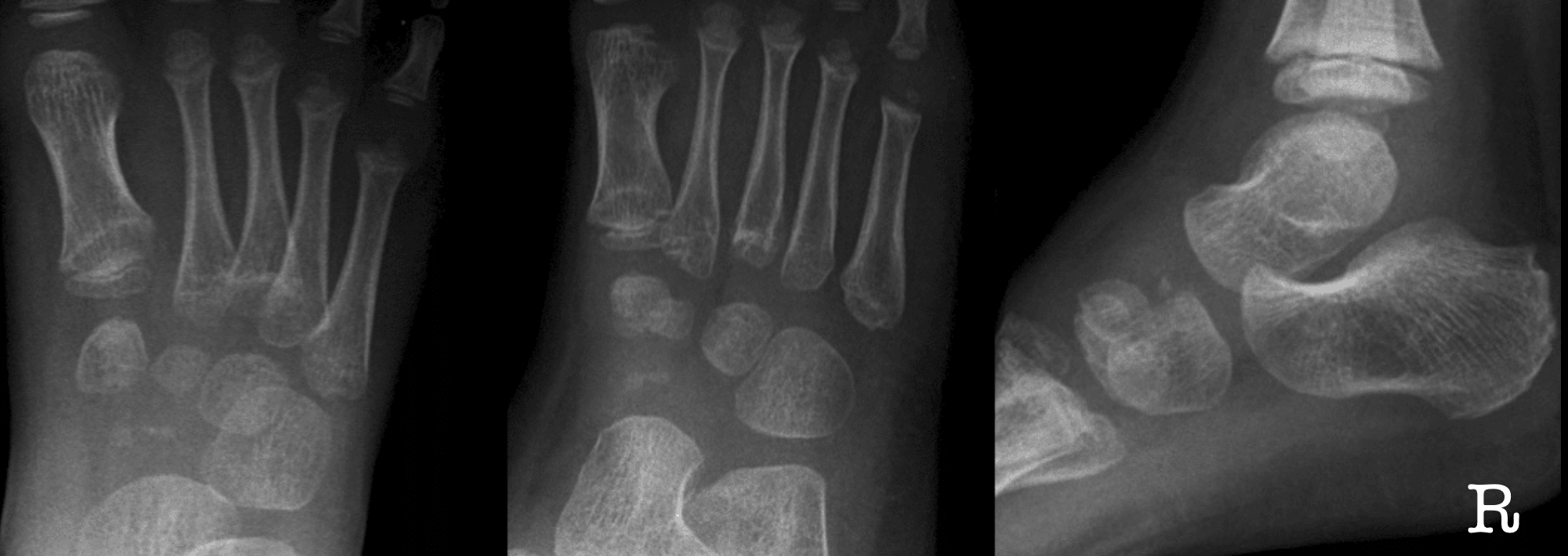
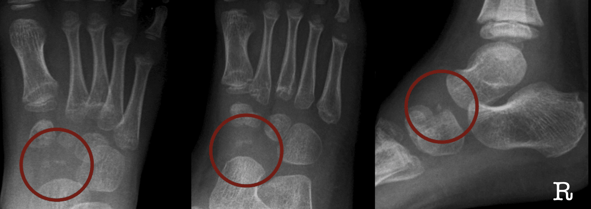
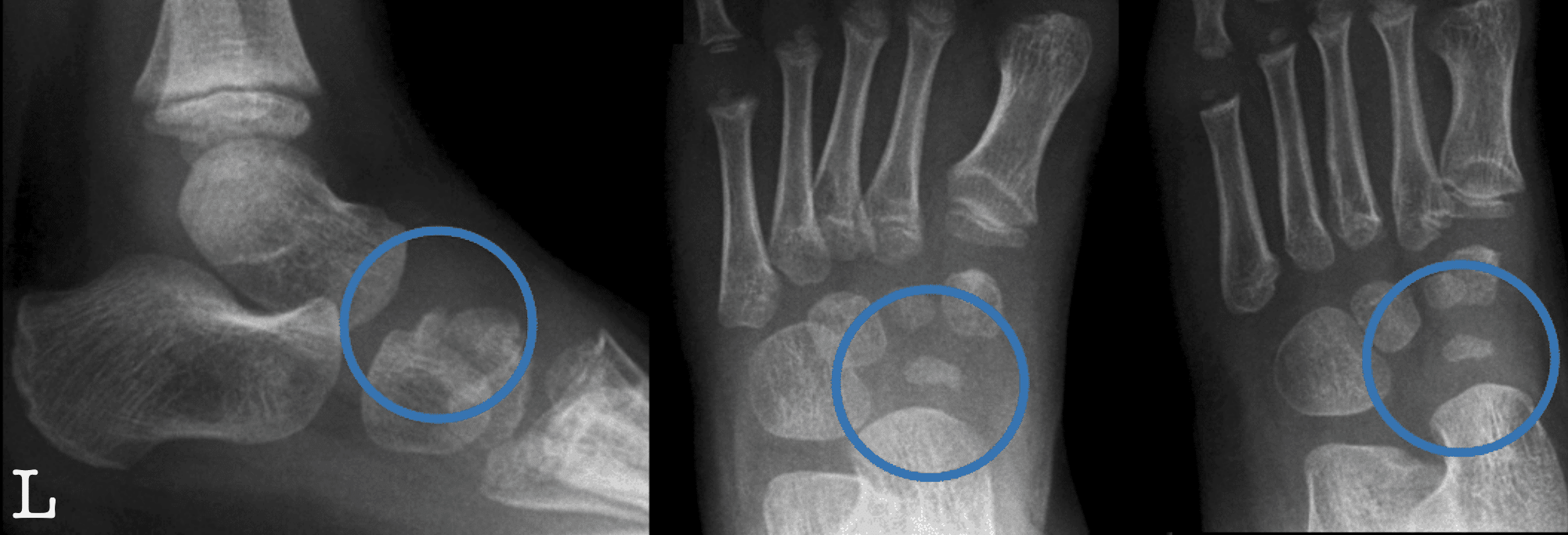
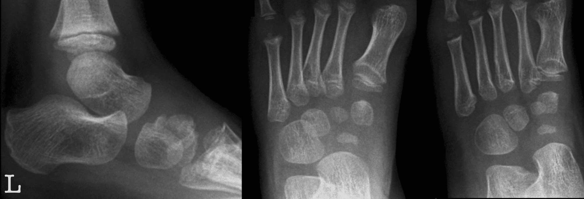
X-ray usually shows a small, flattened or collapsed navicular with increased density, loss of normal trabecular architecture and variable fragmentation. Although these findings look onerous, the natural history is benign, with progressive reossification and restoration of navicular structure.
Management and prognosis
Management is conservative. Analgesia, activity modification and arch support are usually sufficient. A short-leg walking cast for 4–6 weeks may reduce the duration of pain in more symptomatic children. However, immobilisation does not appear to alter the radiographic course or long-term result.
The prognosis is excellent. Long-term studies demonstrate restoration of normal navicular architecture and function without clinically important degenerative sequelae. Persistent or recurrent pain should prompt investigation for an alternative diagnosis such as tarsal coalition or symptomatic accessory navicular.
Differential terminology
Köhler disease is a childhood disorder and should not be confused with Müller-Weiss disease, an adult disorder characterised by progressive deformity and collapse of the tarsal navicular. Note: historical literature may also refer to the tarsal navicular as the tarsal scaphoid.
Case example
Below are images of right foot Navicular avascular necrosis (Köhler disease) in a 5 year old male
History of Köhler disease
1908 – Alban Köhler (1874-1947) first presented the condition at the 4th German Roentgen Congress, then published three cases in Über eine häufige, bisher anscheinend unbekannte Erkrankung einzelner kindlicher Knochen. The patients were boys aged 5–9 years with pain localised to the navicular region. Radiographs showed isolated abnormality of the tarsal navicular while the remaining bones of the foot appeared normal.
Köhler noted “Das Navikulare war in vierfacher Beziehung verändert, und zwar in seiner Grösse, seiner Gestalt, seiner Architektur und seinem Kalkgehalt“. The bone was reduced in size, flattened or irregular, with loss of normal trabecular distinction and increased radiographic density. He observed eventual restoration towards normal and regarded the prognosis as favourable.

Growth disturbance, compression fracture or vascular injury?
1909 – Behn published Isolierte Erkrankung des Naviculare pedis bei Kindern als Zeichen einer Wachstumsstörung interpreting the condition as a disturbance of growth as an isolated occurrence in apparently healthy children. He confirmed the reduction in navicular size, abnormal shape, loss of normal architecture and increased density.
1910 – Kay Schäffer reported a case originally observed in 1907, which he recognised retrospectively after reading Köhler’s reports. The title Die Köhlersche Knochenerkrankung, is the first publication with eponymous attribution.
1911 – Georg Preiser published Zur Frage der typischen traumatischen Ernährungsstörungen der kurzen Hand- und Fusswurzelknochen. He suggested that minor injury could damage the local blood supply, causing rarefaction and central necrosis, with fracture occurring secondarily. He regarded the fracture as less important than the underlying circulatory disturbance and that die Hauptsache müsse eine Gefäss-, bzw. Ernährungsstörung sein! [The principal process must be a vascular, or nutritional, disturbance]
1911 – Stumme published Compressionsfractur des Knochenkerns des Os naviculare pedis proposing compression fracture of the navicular ossification centre as an alternative mechanism. In his 5-year-old patient he interpreted flattening of the nucleus, apparent compression of the trabeculae, a small cortical fragment and approximation of the cuneiform ossification centres towards the talus as evidence of traumatic compression.
Original
English
Alle diese Befunde … lassen unserer Meinung nach nur die eine richtige Erklärung zu, dass es sich hier um eine Kompressionsfraktur des Navikularekerns gehandelt hat.
In our opinion, all these findings permit only one correct explanation: a compression fracture of the navicular ossification centre.
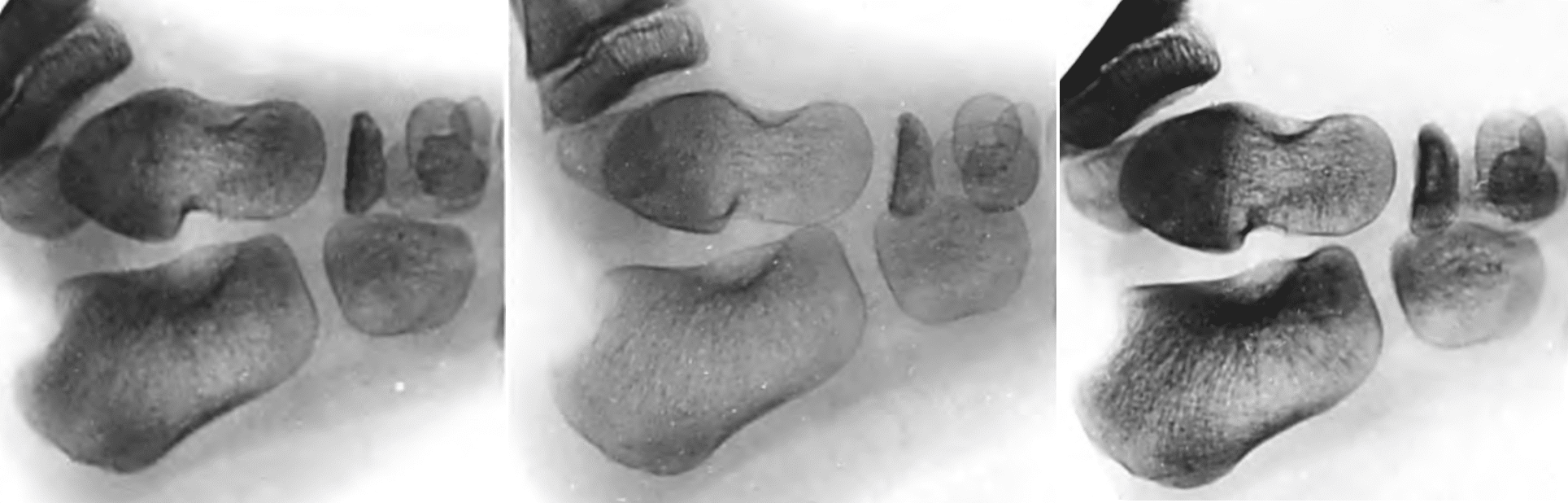
Köhler revisits the pathogenesis
1914 – Köhler presented Zur Pathologie des Os naviculare pedis der Kinde at the 10th German Roentgen Congress. By then he had reviewed 26 published cases. 16 had no history of trauma, four had only trivial trauma, and 10 were bilateral. He rejected the proposition that the radiographic appearance simply represented a fracture or the consequence of a single traumatic event.
I was pleased to see that emphasis on a single trauma, or the view that this was simply a fracture, was regarded in today’s discussion as rather unlikely.
Köhler did not rule out a mechanical influence and proposed that repeated minor mechanical stresses acting on the developing navicular produced a local irritative state, altered vascularity and disturbed ossification. Once this circulatory disturbance resolved, ossification could continue normally.
English-language adoption and recognition of benign natural history
1918 – Charles R. McClure published Isolated Disease of the Scaphoid, noting that the condition first was already known as “Köhler’s disease.” McClure reported on a 7-year-old boy with a small, irregular navicular in which cortex and spongiosa were indistinguishable. After immobilisation, radiographs showed progressive restoration at two months and an almost normal navicular by five months. He considered tuberculosis inconsistent with the rapid and complete recovery and remained unconvinced by trauma as a sufficient explanation.
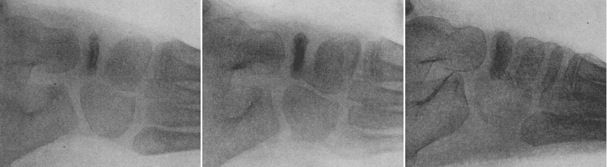
Historical terminology: tarsal scaphoid
Early literature commonly used scaphoid, tarsal scaphoid and os naviculare pedis for the bone now termed the tarsal navicular. At the same time, the modern carpal scaphoid was known as the navicular or os naviculare manus.
Moderny anatomical nomenclature progressively separated the terms, reserving scaphoid for the wrist and navicular for the foot.
1919 – Frederick W. O’Brien published Köhler’s Disease of the Tarsal Scaphoid in Children. He described the condition as a non-infectious process confined to the tarsal scaphoid, with pain, swelling, tenderness on weight bearing, a characteristic radiographic appearance and a good prognosis. O’Brien presented the case of a 3-year-old girl with progressive increase in navicular size and decrease in density over serial radiographs, and clinical recovery after plaster immobilisation.
O’Brien proposed that “the disease appears to be due to delayed development of the ossific centre…”, with trauma acting as a stimulus rather than the primary cause.
1920 – P. Maynard Heath published Isolated Disease of Tarsal Scaphoid-Köhler’s Disease. His case involved a 7-year-old boy with radiographic reduction in navicular size, irregular contour, loss of normal architecture and markedly increased density. Six months later the bone had returned to normal size and the child was asymptomatic.
Heath reviewed the major theories and rejected simple compression fracture and tuberculosis. He considered the disorder primarily an abnormality of ossification, but interestingly argued that it might represent premature rather than delayed ossification. He linked the small dense navicular to weakness of ligamentous support, with secondary strain producing pain and swelling. He suggested the more neutral term “isolated abnormality of the tarsal scaphoid“
1921 – H. Abrahamsen published Koehler’s Disease, Especially with a View to the Pathogeny of the Same. He estimated that about 50 cases had been reported and summarised the principal competing theories:
- fracture of an otherwise normal navicular;
- pathological fracture of a dystrophic navicular;
- delayed formation or primary dystrophic disturbance.
Abrahamsen reported a 7-year-old boy in whom the navicular ossification centre was initially almost absent and then progressively enlarged, eventually acquiring the typical irregular Köhler appearance. He regarded this as an unusually early stage of the process and argued that it supported a developmental abnormality rather than fracture and “the case must be explained as a belated process of ossification.”
1924 – Frederick C. Kidner and Felipe Muro published Köhler’s disease of the tarsal scaphoid or os naviculare pedis retardatum. Their report reviewed 104 cases from 1907 to 1924 and proposed the descriptive term os naviculare pedis retardatum.
They found the disorder most common between 5 and 8 years, more frequent in boys, usually unilateral, and noted that only about one-third of cases had a history of trauma. They therefore considered a single traumatic event unlikely to be the primary cause.
Their case was important because tissue was removed for histology. The specimen showed cellular cartilage with two ossification centres, osteoid septa and imperfect deposition of lime salts, which they interpreted as evidence of interference with normal bone development.
1925 – Frederick E. Diemer and Frank E. Butler published Isolated Disease of the Tarsal Scaphoid (Koehler’s Disease): End-results in Treated and Untreated Cases. They set out to compare functional end-results. They favoured a mechanical-traumatic explanation, arguing that injury to the navicular ossification centres impaired normal bone formation. They proposed the term “osteogenesis imperfecta juvenilis navicularis.
1928 – In the first English edition of Röntgenology: The Borderlands of the Normal and Early Pathological in the Skiagram, Köhler revisited the childhood navicular disorder after two decades of accumulated experience. He rejected a single traumatic fracture as the explanation and warned that fragmentation of the navicular shadow should not itself be mistaken for fracture.
Instead he now favoured a multifactorial mechanical-developmental process:
…the most probable cause is an immoderate strain acting on a normal scaphoid for months at a time or a continuous normal strain acting on a scaphoid weak in development.
Köhler still considered interruption of development the fundamental predisposition, with repeated minor trauma or prolonged loading as contributory factors. He remained sceptical of an inflammatory “scaphoiditis” explanation.
He emphasised the benign natural history with the lesion generally resolving within one to two years with restoration of normal structure, and he advised against operative treatment.
1981 – G. A. Williams and H. R. Cowell published Köhler’s disease of the tarsal navicular, providing one of the first substantial long-term assessments of clinical and radiographic outcome. Williams and Cowell followed 20 patients for an average of approximately 9½ years and found no significant residual alteration in navicular shape at skeletal maturity. Their work helped overturn earlier concern that untreated disease commonly produced permanent navicular deformity and flatfoot.
1984 – Ippolito, Ricciardi Pollini and Falez reported 12 patients followed for an average of 33 years after Köhler disease. All were asymptomatic at final review and none showed degenerative changes attributable to the childhood disorder. Complete restoration of normal navicular bone structure occurred in every patient, taking an average of 8 months.
Nine children had been treated in a weight-bearing plaster cast for three months whilst three received arch supports and rest. Casting relieved pain more rapidly, whereas pain persisted for an average of approximately seven months with arch supports. However, treatment did not alter the ultimate radiographic restoration or long-term result.
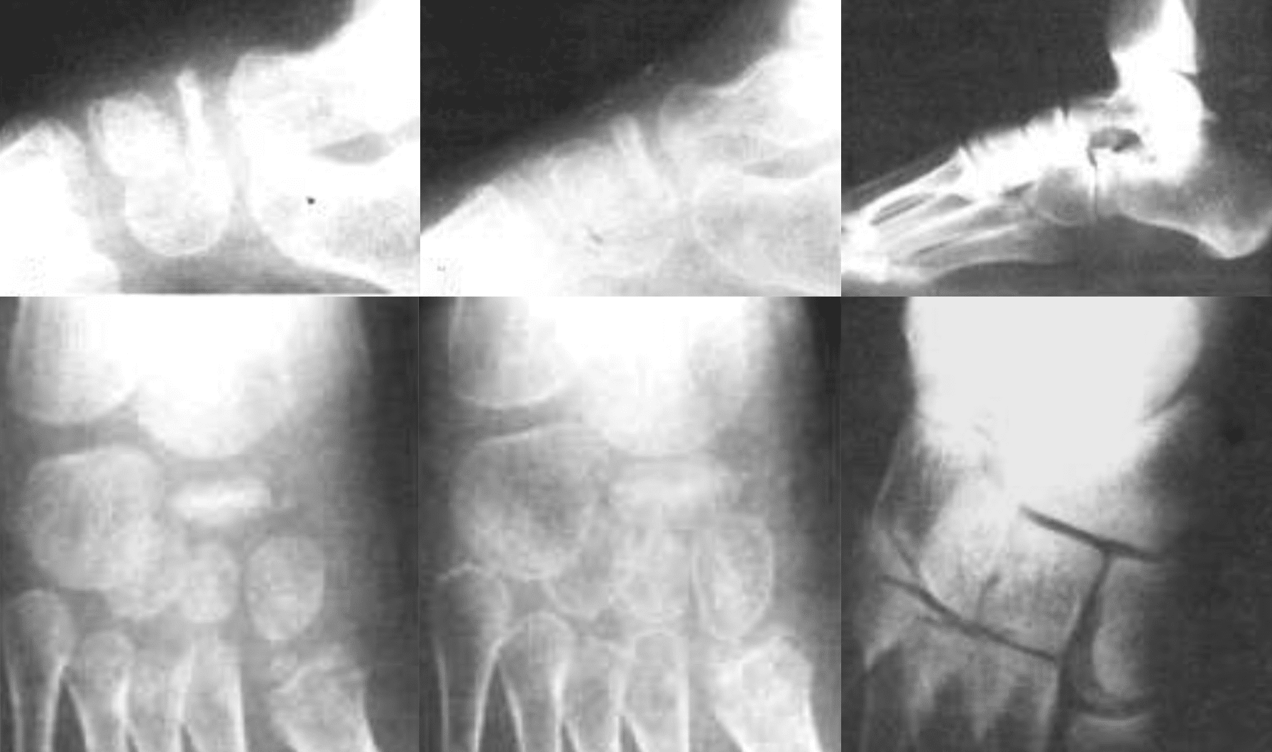
1995 – Jorge Luiz P. Borges et al revisited patients originally treated at the Alfred I. duPont Institute between 1948 and 1975. In their paper Köhler’s bone disease of the tarsal navicular they reviewed fourteen patients with an average follow-up of 31 years 6 months.
The type and duration of treatment did not affect the final clinical or radiographic outcome. However, casting did shorten symptoms. Untreated patients remained symptomatic for an average of 10 months, whereas those treated with a short-leg cast became asymptomatic within an average of 3 months. They concluded that “Köhler’s bone disease is a benign, self-limiting process“
2012 – Shastri, Olson and Fowler summarised Köhler disease as a self-limiting osteonecrosis occurring while the tarsal navicular is still developing. Because the navicular is the last tarsal bone to ossify, it may be compressed between the already ossified talus and cuneiforms, compromising the surrounding vascular supply and producing transient ischaemia of the developing bone.
Associated Persons
- Alban Köhler (1874-1947)
- Walther Müller (1888-1949)
- Konrad Weiss (1891-1976)
- James F Brailsford (1888-1961)
Alternative names
- Kohler disease, Koehler disease
- Osteonecrosis; Osteochondrosis; Osteochondritis of the tarsal navicular
References
Original articles
- Köhler A. Uber eine häufige, bisher anscheinend unbekannte Erkrankung einzelner kindlicher Knochen. Munchener medizinische Wochenschrift, 1908; 55: 1923-1925
- Behn. Isolierte Erkrankung des Naviculare pedis bei Kindern als Zeichen einer Wachstumsstörung. Fortschritte auf dem Gebiete der Röntgenstrahlen. 1909; 14: 262-264
- Schäffer K. Die Köhlersche Knochenerkrankung. Münchener medizinische Wochenschrift. 1910; 57.2(29): .29: 1548-9
- Stumme. Compressionsfractur des Knochenkerns des Os nav. pedis. Fortschritte auf dem Gebiete der Röntgenstrahlen. 1911; 16: 342-346.
- Preiser G. Zur Frage der typischen traumatischen Ernährungsstörungen der kurzen Hand- und Fusswurzelknochen. Fortschritte auf dem Gebiete der Röntgenstrahlen. 1911; 17: 360-362
- Köhler A. Zur Pathologie des Os naviculare pedis der Kinde. Verhandlungen der Deutschen Röntgen-Gesellschaft, 10. Röntgen-Kongress, 1914: 200-203
- Meulengracht. Köhlerske Sygdom i Os naviculare pedis hos Børn. Hospitalstidende. 1915; 58(20): 492-505.
- McClure CR. Isolated Disease of the Scaphoid. JAMA. 1918;71;(17):1360-1361.
- O’Brien F. Köhler’s Disease of the Tarsal Scaphoid in Children. Boston Med Surg J. 1919;180(16):445–447
- Maynard Heath P. Isolated Disease of Tarsal Scaphoid-Köhler’s Disease. Proc R Soc Med. 1920;13(Sect Study Dis Child):17-21.
- Cockayne EA. Isolated Disease of the Scaphoid Bone. Proc R Soc Med. 1920;13(Sect Study Dis Child):41-2.
- Abrahamsen H. Koehler’s Disease, Especially with a View to the Pathogeny of the Same. Acta Radiologica. 1921; 1(2): 174–178
- Kidner FC, Muro F. Köhler’s disease of the tarsal scaphoid or os naviculare pedis retardatum. JAMA 1924;83;(21):1650-1654
- Diemer FE, Butler FE. Isolated Disease of the Tarsal Scaphoid (Koehler’s Disease): End-results in Treated and Untreated Cases. Radiology, 1925; 5(2): 159
- Köhler A. Röntgenology: The Borderlands of the Normal and Early Pathological in the Skiagram. Trans. Arthur Turnbull. William Wood. 1928
Review articles
- Williams GA, Cowell HR. Köhler’s disease of the tarsal navicular. Clin Orthop Relat Res. 1981 Jul-Aug;(158):53-8.
- Ippolito E, Ricciardi Pollini PT, Falez’ F. Köhler’s disease of the tarsal navicular: long-term follow-up of 12 cases. J Pediatr Orthop. 1984; 4(4): 416-7
- Borges JL, Guille JT, Bowen JR. Köhler’s bone disease of the tarsal navicular. J Pediatr Orthop. 1995 Sep-Oct;15(5):596-8.
- Shastri N, Olson L, Fowler M. Kohler’s Disease. West J Emerg Med. 2012; 13(1): 119–120.
- Cadogan M. Eponymythology of foot injuries. LITFL
- Cadogan M. Carpal and tarsal bone nomenclature. LITFL
eponymictionary
the names behind the name

Sujit Iyer, MD, is the National Director of Pediatric Services at US Acute Care Solutions and a Professor of Pediatrics and Pediatric EM Fellowship Director at UT Austin Dell Medical School. He completed his medical training and pediatrics residency at Baylor College of Medicine / Texas Children’s Hospital, followed by a Pediatric Emergency Medicine fellowship at Children’s Hospital of Philadelphia. Dr. Iyer focuses on emergency department operations, pediatric readiness in community hospitals, and curriculum development, lecturing nationally on quality improvement and pediatric emergency medicine best practices.
BA MA (Oxon) MBChB (Edin) FACEM FFSEM. Emergency physician, Sir Charles Gairdner Hospital. Passion for rugby, medical history, medical education, and asynchronous learning #FOAMed evangelist. Editor-in-Chief and Webmaster of Life in the Fast lane, LITFL | On Call 4e| Eponyms | Books | Horology |