
![]()
Jan Evangelista Purkinje
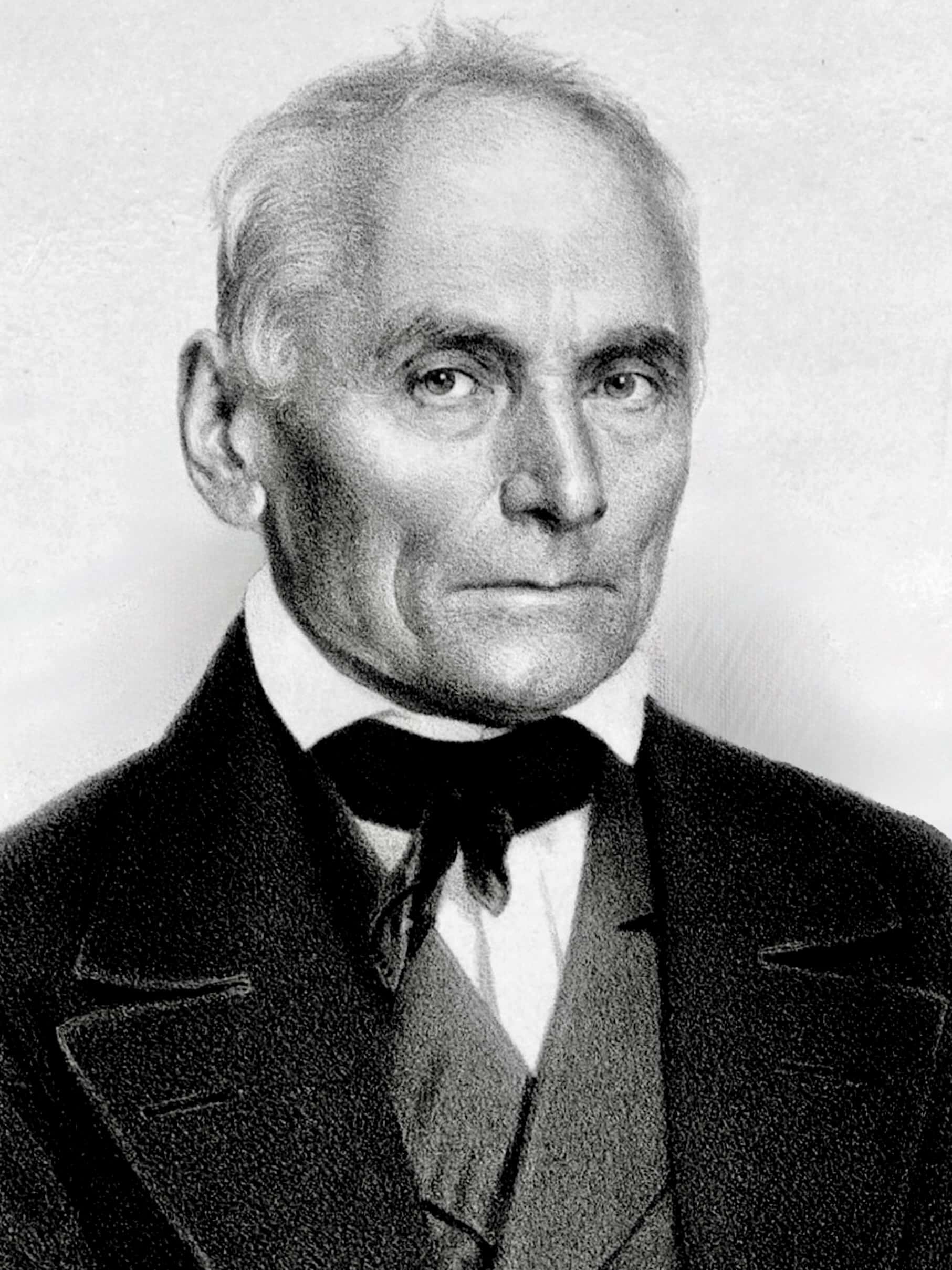
Jan Evangelista Purkyně (1787-1869) was a Czech anatomist and experimental physiologist
Purkyně was one of the most prolific and wide-ranging physiologists of the 19th century, whose name is immortalised across disciplines from neuroscience and cardiology to ophthalmology and dermatology. Trained in medicine at the University of Prague and later appointed professor of physiology in Breslau, Purkyně combined meticulous self-experimentation with pioneering use of microscopy. His investigations opened new fields of study, describing cellular structures, tissue organisation, and physiological processes with a precision far ahead of his time.
In vision science, Purkyně’s work laid foundational stones for modern ophthalmology and neurophysiology of sight. He documented reflections from the cornea and lens (Purkinje images); explored afterimages, entoptic phenomena, the Purkinje shift in spectral sensitivity, and complex subjective visual experiences such as the Purkinje tree of the retinal vessels.
In neuroanatomy, he identified the large cerebellar neurons the Purkinje cells and described their arborisation. In cardiology, he discovered the Purkinje fibres of the ventricular conduction system, elucidating their role in coordinating heartbeat. His observations on ciliary motion in epithelial tissues revealed a ubiquitous mechanism of cellular motility. He even investigated the microstructure of skin, fingerprints, and other physiological systems, producing original insights that remain relevant in today’s medical science.
Biography
- Born December 17, 1787 in Libochovice, Bohemia (Austrian Empire; now Czech Republic)
- 1793–1796 Primary schooling in Libochovice.
- 1798–1804 Choirboy at Piarist monastery in Mikulov; Entered Piarist order at Stará Voda monastery (1804); received title “Professor in Humanibus Litteris”.
- 1806 – Studied at Piarist Philosophical Institute, Litomyšl; qualified for university entry.
- 1807–1809 Student, Philosophical Institute, University of Prague; mainly physical sciences.
- 1809–1812 Private tutor to Baron Hildprandt’s son.
- 1812 -1818 Medical studies at University of Prague. Defended MD dissertation Beiträge zur Kenntniss des Sehens in subjektiver Hinsicht; Received MD degree (1818).
- 1819–1822 Instructor in anatomy, Prague Medical Faculty.
- 1823 – Appointed Professor of Physiology & Pathology, University of Breslau. Habilitation dissertation: Commentary on the Physiological Examination of the Visual Organs and the Cutaneous System; Published first classification of fingerprints.
- 1825 – Published Neue Beiträge zur Kenntniss des Sehens in subjektiver Hinsicht (New Subjective Reports about Vision).
- 1833 – Discovered sweat glands.
- 1837 – Described Purkinje cells in cerebellum.
- 1839 – Discovered Purkinje fibres in heart; founded first physiological institute, Breslau.
- 1842 – Developed laboratory-based teaching of physiology.
- 1850 – Professor of Physiology, University of Prague; promoted Czech language teaching.
- 1853 – Co-founded Živa science periodical.
- 1861 – Co-founded Society of Czech Physicians and Časopis lékařů českých; elected deputy member, Bohemian Provincial Diet (1861–1866).
- 1868 – Awarded Imperial Austrian Order of Leopold.
- Died July 31, 1869 in Prague after prolonged illness. Buried with honours at Vyšehrad National Cemetery, Prague.
Medical Eponyms
Purkinje Cells (1837)
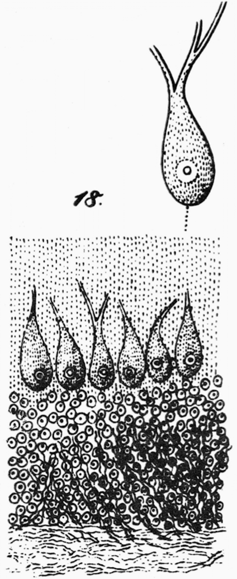
Purkinje cells are inhibitory GABAergic neurons forming the sole output of the cerebellar cortex. Their dendrites receive synaptic input from parallel fibres of granule cells; their axons project to the deep cerebellar nuclei.
Purkyně’s 1837 presentation to the Gesellschaft Deutscher Naturforscher und Aerzte is the earliest printed account of the cerebellar neurons that now bear his name. He described large, flask-shaped neurons in the cerebellar cortex with extensive dendritic arborisation. These large, flask-shaped cells form a single layer between the molecular and granular layers, they integrate massive synaptic input and send inhibitory projections to the deep cerebellar nuclei.
Purkyně’s observation of their orientation, branching processes, and distinct location foreshadowed modern neuroanatomical mapping of neuronal circuits.
Each of these bodies is directed inward toward the cerebrum with its blunt, rounded end, and clearly shows the central nucleus within its bulb and its halo. The other, tail-shaped end is directed outward and, with usually two processes, disappears into the grey matter near the outer periphery, where it is encased in the vascular membrane. (Fig. 18.)
Purkinje 1837
Purkinje Fibres (1839)
Purkinje fibres are specialised myocardial fibres with high conduction velocity, part of the His–Purkinje network and essential for synchronised ventricular depolarisation.
In 1839, Purkyně introduced the concept of morphologically distinct myocardial tissue, suggesting structural specialization in the heart. He provided the first published account of what we now call Purkinje fibres, specialized muscle fibres forming part of the ventricular conduction system.
Located subendocardially, these fibres are larger, lighter staining, and contain fewer myofibrils than working myocardium, enabling rapid conduction of electrical impulses from the atrioventricular node to the ventricular myocardium for coordinated contraction.
Original
English
W ścianach komór serca dostrzegłem szczególne, jasne i błyszczące włókna, odróżniające się od mięśnia sercowego swym wyglądem i układem, tworzące siatkowatą sieć biegnącą pod wsierdziem
In the walls of the heart’s ventricles I observed peculiar, pale and glistening fibres, differing from the cardiac muscle in appearance and arrangement, forming a reticular network running beneath the endocardium – Purkyniego 1839
Purkinje vesicle (1825)
While examining unfertilised hen’s eggs, Purkyně identified a small, clear, spherical body within the yolk, naming it the vesicula germinativa. He correctly interpreted it as the nucleus of the ovum, making this one of the earliest clear descriptions of a nucleus in animal tissue.
…in vitello ovi avium globulum pellucidum observavi, quem vesiculam germinativam nominavi…
Symbolae ad ovi avium historiam ante incubationem
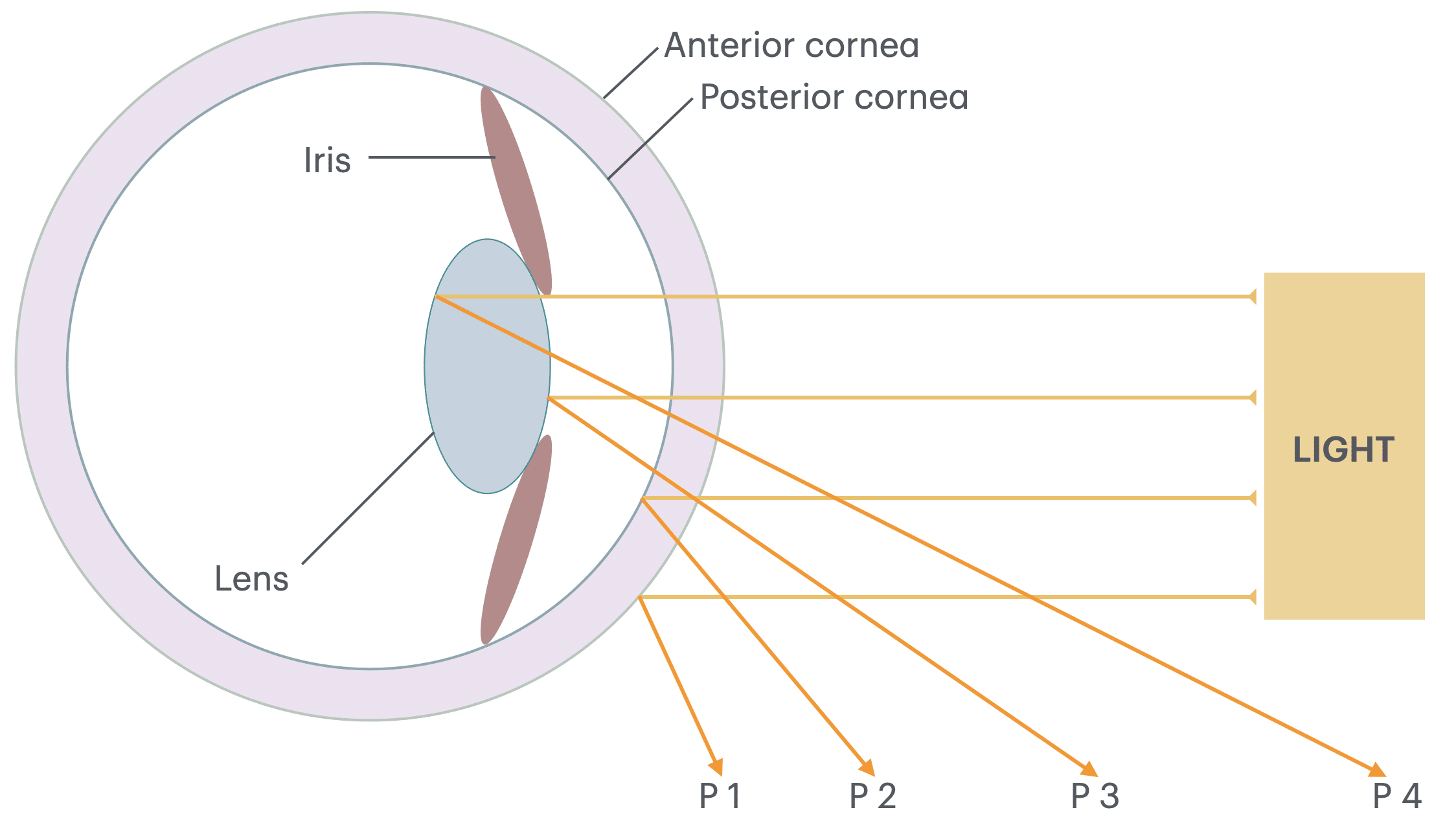
Purkinje Images (1823)
Purkinje images are reflections of light (objects) from ocular surfaces first described by Purkyně in 1823. Also known as Purkinje reflexes or Purkinje–Sanson images.
- (P1) reflection from the outer (anterior) surface of the cornea (erect, bright, distinct)
- (P2) reflection from the inner (posterior) surface of the cornea (erect, faint)
- (P3) reflection from the outer (anterior) surface of the lens (erect, faint, smaller)
- (P4) reflection from the inner (posterior) surface of the lens (inverted, faintest, smallest)
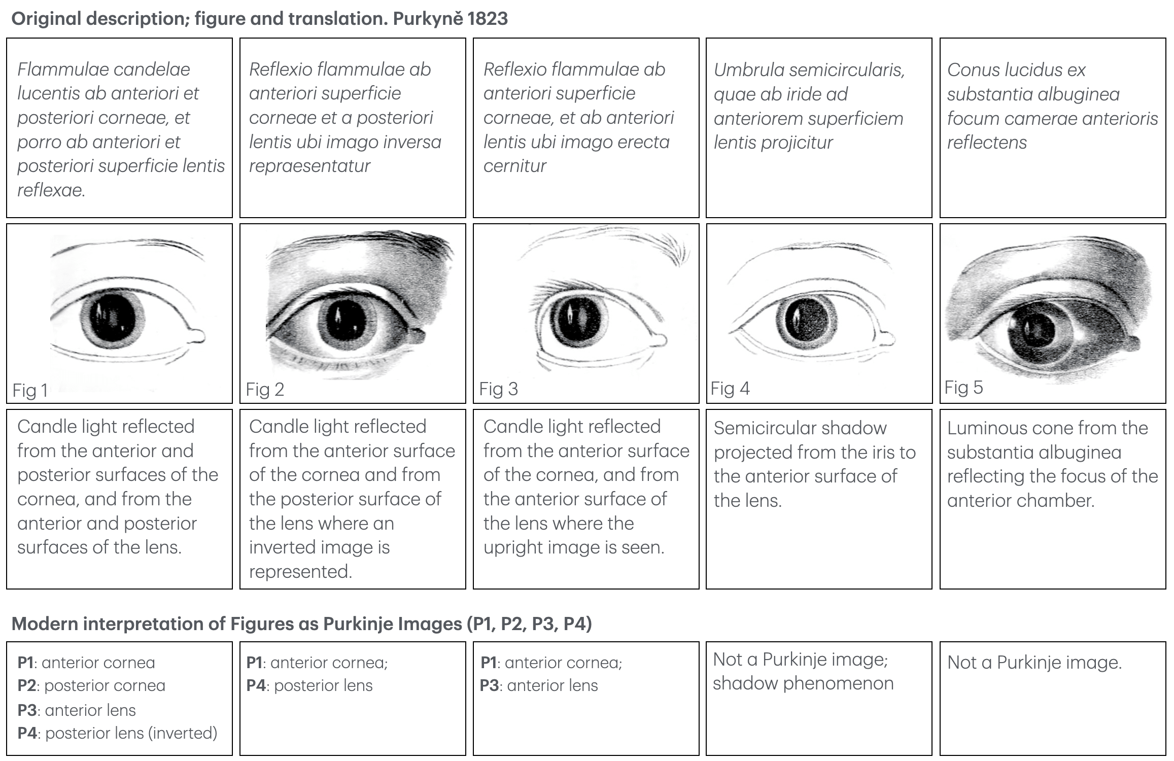
Today these are known as the first, second, third and fourth Purkinje images. Modern numbering (P1–P4) was formalised to align optical and anatomical conventions. The images are used in optical research, used to study accommodation, eye movements, and ocular biometry.
P1 – Anterior cornea: erect, bright, largest image
P2 – Posterior cornea: erect, smaller, dimmer
P3 – Anterior lens: erect, faint, smaller
P4 – Posterior lens: inverted, faintest, smallest
Purkinje Shift (1825)
The Purkinje shift (Purkinje phenomenon) describes the change in the eye’s peak spectral sensitivity of the human eye from ~555 nm (green-yellow, cone-mediated photopic vision) in bright light to ~507 nm (blue-green, rod-mediated scotopic vision) in dim light. This accounts for the relative darkening of reds and brightening of blues as illumination falls.
Purkyně first documented the phenomenon in 1825 in Neue Beiträge zur Kenntniss des Sehens in subjectiver Hinsicht. He noticed that the apparent brightness of colours changes with illumination level — reds becoming darker and blues/greens relatively brighter in dim light.
Original
English
Abends bemerkte ich, daß die rothen Blumen, die ich bei Tage am hellsten gesehen hatte, nun schwärzer erschienen, während die blauen Blumen verhältnißmäßig heller leuchteten.
In the evening I noticed that the red flowers, which in the daytime I had seen as the brightest, now appeared darker, while the blue flowers shone relatively brighter. – Purkinje 1825: 110
Purkyně’s observation was made decades before the physiological distinction between rods and cones was known, yet it remains a foundational description of human visual adaptation and is still referenced in vision science and lighting design today.
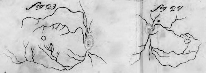
Purkinje Tree (1823)
Purkinje first described the phenomenon in Beobachtungen und Versuche zur Physiologie der Sinne (1823) while exploring subjective visual effects. By illuminating the eye in a particular way (often obliquely with a candle or sunlight), he could perceive the branching pattern of his own retinal blood vessels projected into his field of vision. This tree-like network (Purkinje tree), is an entoptic image created when light casts shadows of the retinal vessels onto photoreceptor cells.
Fig. 23 – Detailed depiction of the retinal vasculature seen by Purkinje.
Fig. 24 – Variant of the vascular pattern.
Original
English
Wenn ich das Auge vor einer Lichtquelle bewege, so erscheint ein dunkler, verästelter Schatten, der mit meinen Augenbewegungen sich bewegt, und den ich für die Adern der Netzhaut halte
Under certain conditions of illumination, one can perceive the image of the blood vessels of the retina itself, in a network resembling a tree that spreads over the entire field of vision – Purkinje 1823: 19
The Purkinje tree is an entoptic phenomenon caused by shadows from the retinal blood vessels, most visible when the eye is illuminated by a moving point source close to the sclera. This movement shifts the image on the retina, stimulating photoreceptors in areas normally shielded by blood vessels, making them visible. It is often demonstrated in vision science teaching using a bright LED or ophthalmoscope light.
Purkinje Afterimages (1823)
Purkyně’s fascination with subjektive Erscheinungen (subjective phenomena) led him to a detailed study of afterimages. In the chapter Das Nachbild. Imagination , Gedächtnits des Gesichtssinnes, he explores their forms, duration, and transformations, anticipating many modern insights into sensory adaptation.
Original
English
Das Nachbild ist das Fortdauern des Bildes eines Gegenstandes im Gesichtssinne, nachdem derselbe dem Auge entzogen worden ist…Bei starkem Lichte erscheint das erste Nachbild gleichgefärbt mit dem wirklichen Gegenstande; später verändert es sich in die entgegengesetzte Farbe…e länger das Auge ermüdet ist, desto schneller tritt die Umwandlung in die Complementärfarbe ein.
The afterimage is the persistence of the image of an object in the sense of vision after it has been withdrawn from the eye. In strong light the first afterimage appears the same colour as the actual object; later it changes to the opposite colour…The more fatigued the eye is, the more quickly the transformation into the complementary colour occurs. – Purkinje, 1823
Afterimages occur because of photoreceptor bleaching and neural adaptation. Positive afterimages reflect continued activity of the same retinal receptors that were stimulated by the original image; negative afterimages occur when these receptors are fatigued and the visual system shifts perception toward the complementary colour.
Purkyně’s meticulous self-observations anticipated experimental psychophysics, and his combination of colour, time, and intensity variables foreshadowed later studies by Helmholtz and Hering.
Key Medical Contributions
Ciliary Motion (Purkyně & Valentin, 1834–1835)
In early 1834, Gabriel Gustav Valentin, working with Purkyně in Breslau, observed rhythmic motion in the epithelial lining of a rabbit’s oviduct. Purkyně recognised this as an example of ciliary motion, recalling similar activity in aquatic animals he had previously studied.
Their joint 1834 German paper publicly announced the discovery of continuous ciliary movement in vertebrates, and their 1835 Wratislavia Latin treatise presented a comprehensive experimental study. They documented the presence of cilia in numerous organs, tested 55 chemical and physical agents, and demonstrated that ciliary motion is an autonomous property of the cilia, unaffected by nervous or muscular control.
Original
English
Wir haben eine neue, bisher unbekannte Bewegung entdeckt, welche durch Wimperhaare hervorgebracht wird, und als ein allgemeines Phänomen in den Klassen der Amphibien, Vögel und Säugethiere vorkommt [1834]
…motus vibratorius iste neque a voluntate neque a nervorum influxu pendet, sed vi quadam propria partibus inhaerente continenter producitur…[1835]
We have discovered a new, hitherto unknown movement, produced by cilia, and occurring as a general phenomenon in the classes of Amphibia, Birds, and Mammals.
…this vibratory motion depends neither on the will nor on the influence of the nerves, but is continuously produced by a certain force inherent in the parts themselves… Purkinje, Valentin 1834.
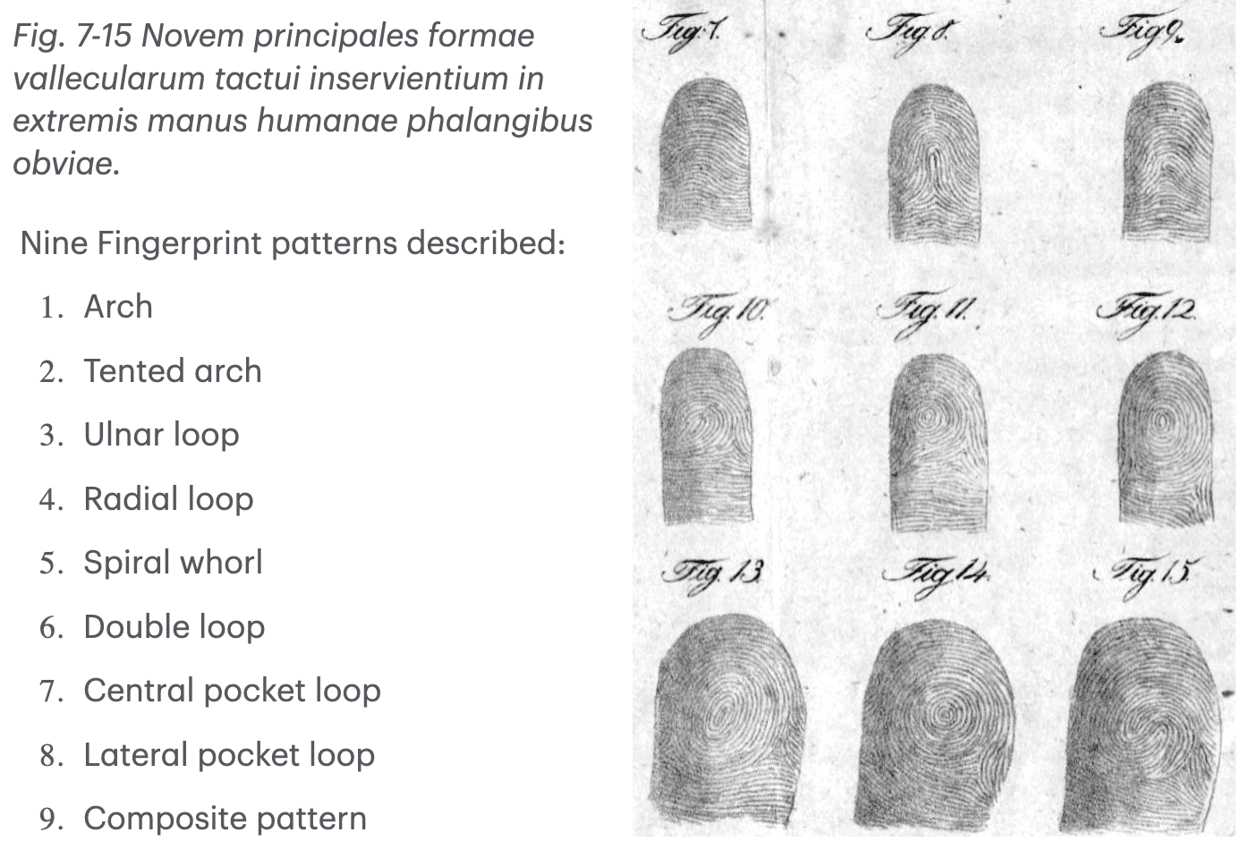
Purkinje and Fingerprints (1823)
No systematic taxonomy for fingerprints existed before Purkyně. Fingerprints appear on Babylonian clay tablets (~2000 BCE), Chinese seals, and Persian pottery, used for authentication, not scientific study.
Early anatomical notes were provided by Nehemiah Grew (1648–1712) who described ridge patterns in Philosophical Transactions (1684); Govard Bidloo (1649–1713) included fingertip ridge drawings in Anatomia Humani Corporis (1685); and Marcello Malpighi (1628–1694) described ridges but did not classify them in his Opera Omnia (1686).
In his Latin monograph Commentatio de examine physiologico organi visus et systematis cutanei, Purkyně examined the structure, sensitivity, sweat pores, and papillary ridges of the skin. He systematically classified fingerprint ridge patterns into nine geometric types, with engraved illustrations marking the first scientific classification in history.
„…in digitorum apicibus atque in palmis ac plantis cutis eminentiae papillaris in certas figuras dispositas conspiciuntur….“
“…on the tips of the fingers and on the palms and soles, the papillary elevations of the skin are arranged in certain patterns…”
Purkyně’s work was purely anatomical and physiological, not forensic. His meticulous ridge classification anticipated the dermatoglyphic studies of the 20th century and the forensic applications of the late 19th century.
Self-Experimentation in Psychopharmacology
Purkyně was not only a pioneer in experimental physiology, but also an early and fearless self-experimenter in pharmacology. Decades before Buchheim founded modern experimental pharmacology, Purkyně investigated drug effects by using himself as the subject, documenting both objective signs and subjective sensations that animal experiments could not reveal.
In 1825, in Beobachtungen und Versuche zur Physiologie der Sinne he published detailed accounts of belladonna’s ocular effects including mydriasis, chromatic “stellate” figures, and altered light dispersion, attributing them to lens changes from cycloplegia. His digitalis experiments described unique “flicker roses” in vision, linked to vagus-mediated effects via nausea rather than a direct retinal action.
Purkyně’s 1829 Einige Beiträge zur physiologischen Pharmacologie extended to camphor (producing euphoria and heightened spirituality), belladonna and stramonium (secretory inhibition, pupil dilation), turpentine and nutmeg (hypnotic and narcotic effects, potentiated by wine), and early recognition of drug–drug interactions, now seen as potentiation or sensitisation. He and his student Valentin also tested 55 agents on ciliary motion (1834), deducing functional independence from the CNS.
While his method was inherently risky, Purkyně’s meticulous documentation prefigured modern human pharmacology and offered enduring insights into drug action, subjective experience, and physiological mechanisms.
Controversies
Did Purkyně Invent the Ophthalmoscope?
Claims that Purkyně invented the ophthalmoscope appear frequently in secondary literature and online sources. The origin of this claim lies in his 1823 candlelight experiments, which shared some optical principles later used by Helmholtz, but were fundamentally different in purpose and outcome.
Purkyně’s 1823 experiment: (Beobachtungen und Versuche zur Physiologie der Sinne, p. 48)
„Ich stellte das Licht einer Kerze seitwärts vor das Auge und beobachtete die Spiegelbilder, welche von den verschiedenen Flächen desselben zurückgeworfen werden.“
“I placed the light of a candle to the side of the eye and observed the mirror images reflected from its various surfaces.”
This work described the Purkinje images as reflections from the anterior/posterior cornea and anterior/posterior lens surfaces of a candle flame. These studies involved observing reflections, not the interior of the living retina.
In 1851, Hermann von Helmholtz (1821-1894) introduced a device combining a light source and a semitransparent mirror (or perforated reflector) aligned with the observer’s eye, allowing direct visualisation of the retina in vivo. He acknowledged prior optical studies of the eye but did not attribute the ophthalmoscope’s invention to Purkyně.
Source of confusion:
- Purkyně’s experiments used external light alignment and observation along nearly the same axis the same principle that makes ophthalmoscopy possible.
- Later popular accounts and obituaries romanticised this connection, labelling Purkyně’s setup a “primitive ophthalmoscope.”
- Historical ophthalmology texts (late 19th century) sometimes credited him with “anticipating” the ophthalmoscope, which gradually morphed into “inventing” it in some non-academic sources.
Purkyně should be credited with early demonstrations of ocular retroillumination and the discovery of the Purkinje images, but not with the invention of the ophthalmoscope. The instrument itself, which enabled detailed retinal examination, was the work of Helmholtz in 1851.
Inconsistencies
Birth date: Most modern references give December 17, 1787 (e.g., Czech biographical sources), while others list December 18, 1787. No definitive contemporary birth or baptism record has yet resolved the discrepancy.
Goethe’s role in Breslau appointment: Some narratives state that Johann Wolfgang von Goethe personally intervened to secure Purkyně’s appointment to the University of Breslau. Others note the absence of direct evidence and treat the claim as anecdotal.
Purkinje fibres – disputed first publication: Purkyně’s earliest verifiable description of the fibres is in Polish (Nowe spostrzeżenia i badania w przedmiocie fizjologii i drobnowidowej anatomii, 1839). However, numerous secondary sources cite a Latin pamphlet entitled Symbolae ad anatomiam fasciculi nervosi auriculi cordis as the first publication, but no catalogue entry, archival holding, or digitised copy has been found. This suggests the title may be apocryphal or a misattribution.
Names and transliterations: Published under multiple linguistic forms with name changes including:
- Forename: Jan, Johann, and Joannes.
- Middle name: Evangelist or Evangelista.
- Surname: Purkyně, Purkinje, Purkyne, Purkinieg, Purkiniego
Major Publications
- Purkinje JE. Beiträge zur Kenntniss des Sehens in subjectiver Hinsicht. Prag: Calve, 1819
- Purkinje JE. Beiträge zur Kenntniss des Sehens in subjectiver Hinsicht Berlin: Reimer, 1823 (2e)
- Purkinje JE. Beobachtungen und Versuche zur Physiologie der Sinne. Neue Beiträge zur Kenntniss des Sehens in subjectiver Hinsicht. Berlin: Reimer. 1825
- Purkyně J.E. Commentatio de examine physiologico organi visus et systematis cutanei : quam pro loco in gratioso medicorum ordine rite obtinendo die XXII. Wratislaviae: Korn, 1823. [Fingerprints]
- Purkinje JE. Symbolae ad ovi avium historiam ante incubationem. 1825 [Purkinje vesicle]
- Purkinje JE. Einige Beiträge zur physiologischen Pharmacologie. Neue Breslauer Sammlungen aus dem Gebiete der Heilkunde 1829; 1: 423-43
- Purkinje JE. Der microtomische Quetscher, ein bei microscopischen Untersuchungen unentbehrliches Instrument. Archiv für Anatomie, Physiologie und wissenschaftliche Medicin 1834: 385–390.
- Purkinje JE, Valentin G. Entdeckung continuirlicher durch Wimperhaare erzeugter Flimmerbewegungen, als eines allgemeinen Phänomens in den Klassen der Amphibien, Vögel und Säugethiere. Archiv für Anatomie, Physiologie und wissenschaftliche Medicin 1834; 391-400 [Ciliary Motion]
- Purkinje JE, Valentin G. De phaenomeno generali et fundamentali motus vibratorii continui in membranis cum externis tum internis animalium plurimorum et superiorum et inferiorum ordinum obvii : commentatio physiologica. Wratislaviae, 1835 [Ciliary Motion]
- Purkinje JE. Section 16. Neueste Untersuchungen aus der Nerven- und Hirnanatomie In: Amtlicher Bericht über die Versammlung der Gesellschaft Deutscher Naturforscher und Aerzte (Prag), 1837: 177–180, Fig. 17 [Purkinje Cells]
- Purkinje JE. Nowe spostrzeżenia i badania w przedmiocie fizjologii i drobnowidowej anatomii udzielane przez naszego korespondenta Dr. J. E. Purkyniego p.z. profesora Fizjologii i Patologii we Wrocławiu. Rocznik Wydziału Lekarskiego w Uniwersytecie Jagiellońskim. Vol. 1839;II:44–67 [Purkinje Fibres]
- Purkinje JE. Mikroscopisch-neurologische Beobachtungen. Archiv für Anatomie, Physiologie und wissenschaftliche Medicin 1845; 12: 281–295.
- Purkinje JE. Commentatio de examine physiologico organi me el systematis cutane In: Sebrané spisy
References
Biography
- John HJ. Jan Evangelista Purkyně Czech Scientist and Patriot (1787–1869). Proceedings of the Royal Society of Medicine. 1953; 46(11): 933-940.
- John HJ. Jan Evangelista Purkyne Czech Scientist and Patriot 1787-1869. 1959
- Haas LF. Jan Evangelista Purkinje (1787-1869). J Neurol Neurosurg Psychiatry. 1994 Jul;57(7):777
- Davies MK, Hollman A. Jan Evangelista Purkinje (1787-1869). Heart. 1996 Oct;76(4):311.
- O’Connor C. Jan Evangelista Purkyne – a groundbreaking scientist who played a major role in the Czech national revival. 2006
- Živa: Special Issue on J. E. Purkyně, No. 5, 2011. (Czech)
- Waliszewska-Prosół M, Ejma M, Podemski R. Jan Ewangelista Purkynie (1787-1869) – fizjolog, fenomenolog, wrocławianin [Jan Ewangelista Purkynie (1787-1869) – physiologist, phenomenologist, citizen of Wroclaw]. Neurol Neurochir Pol. 2013 Jan-Feb;47(1):90-3.
- Grzybowski A, Pietrzak K. Jan Evangelista Purkynje (1787-1869). J Neurol. 2014 Oct;261(10):2048-50.
- Mazurak M, Kusa J. Jan Evangelista Purkinje: A Passion for Discovery. Tex Heart Inst J. 2018; 45(1): 23-26.
- Fresquet JL. Johannes Evangelista Purkinje (1787-1869). Historia de a la Medicina
- Jan Evangelista Purkyně. Monoskop
- Minai M. Jan Evangelista Purkyne (1787-1869). Embryo Project
Eponymous terms
Self-Experimentation
- Hanzlik PJ. Purkinje’s Pioneer Self-Experiments in Psychopharmacology: Part I. Cal West Med. 1938 Jul;49(1):52-5
- Hanzlik PJ. Purkinje’s Pioneer Self-Experiments in Psychopharmacology: Part II. Cal West Med. 1938 Aug;49(2):140-2.
Eyes, vision, images and the ophthalmoscope
- Bowman W. Lectures on the parts concerned in the operations on the eye, and on the structure of the retina : delivered at the Royal London Ophthalmic Hospital, Moorfields, June 1847: 59-60
- Sherman SE. The history of the ophthalmoscope. Doc Ophthalmol 1989; 71, 221–228 (1989)
- Reese PD. The neglect of Purkinje’s technique of ophthalmoscopy prior to Helmholtz’s invention of the ophthalmoscope. Ophthalmology. 1986 Nov;93(11):1457-60.
Cells, physiology and neuroscience
- Teich M. Purkyně and Valentin on Ciliary Motion: An Early Investigation in Morphological Physiology. The British Journal for the History of Science. 1970;5(2):168-177
- Tan SY, Lin KH. Johannes Evangelista Purkinje (1787-1869): 19th century’s foremost phenomenologist. Singapore Med J. 2005 May;46(5):208-9.
- Zárský V. Jan Evangelista Purkyně/Purkinje (1787-1869) and the establishment of cellular physiology–Wrocław/Breslau as a central European cradle for a new science. Protoplasma. 2012 Oct;249(4):1173-9.
- Cavero I, Guillon JM, Holzgrefe HH. Reminiscing about Jan Evangelista Purkinje: a pioneer of modern experimental physiology. Adv Physiol Educ. 2017; 41(4): 528-538.
- Chvátal A. Jan Evangelista Purkyně (1787–1869) and his instruments for microscopic research in the field of neuroscience. Journal of the History of the Neurosciences, 2017; 26(3): 238–256.
- Buttner R, Lee J. De-eponymising anatomical terminology. LITFL
Fingerprints
- Grew N. The description and use of the pores in the skin of the bands and feet. Phil. Trans. R. Soc. 1684; 14: 566–567
- Bidloo G. Anatomia Humani Corporis. 1685.
- Malpighi M. Opera Omnia. 1686.
- Cummins H, Kennedy RW. Commentatio de examine physiologico organi visus et systematis cutanei. Purkinje’s observations (1823) on finger prints and other skin features. 1940. Birth Defects Orig Artic Ser. 1991;27(2):19-64.
- Grzybowski A, Pietrzak K. Jan Evangelista Purkynje (1787-1869): first to describe fingerprints. Clin Dermatol. 2015 Jan-Feb;33(1):117-21.
Legacy
Purkyně’s legacy is as expansive as his curiosity. His name is etched into anatomical nomenclature and physiological vocabulary across multiple branches of medicine, yet his influence runs deeper than eponyms. He helped professionalise physiology as an independent academic discipline, established one of Europe’s first dedicated physiological institutes, and mentored a generation of scientists who carried his experimental ethos forward.
By integrating fine microscopy with careful experimentation and self-observation, he bridged the descriptive tradition of anatomy with the quantitative approach of modern biomedical science. Two centuries after his birth, Purkyně’s discoveries continue to be taught in medical schools worldwide, a testament to the enduring power of meticulous observation and intellectual range.
Eponym
the person behind the name
BA MA (Oxon) MBChB (Edin) FACEM FFSEM. Emergency physician, Sir Charles Gairdner Hospital. Passion for rugby; medical history; medical education; and asynchronous learning #FOAMed evangelist. Co-founder and CTO of Life in the Fast lane | On Call: Principles and Protocol 4e| Eponyms | Books |