
![]()
Friedrich Horner
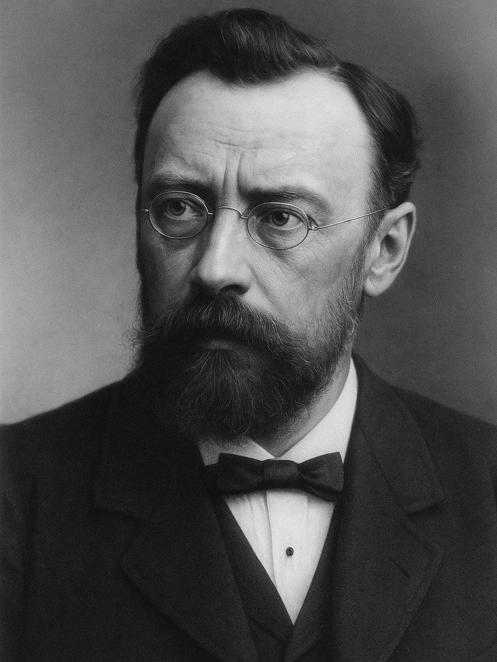
Johann Friedrich Horner (1831-1886) was a Swiss ophthalmologist.
Friedrich Horner was an ophthalmologist and academic, widely regarded as the founder of modern ophthalmology in Switzerland. He was instrumental in introducing antiseptic principles into ophthalmic surgery in Switzerland and was an early proponent of rigorous aseptic techniques. He published extensively on conditions such as glaucoma, retinal detachment, and lacrimal system diseases. His emphasis on anatomical precision and cleanliness contributed significantly to surgical outcomes in the pre-antibiotic era.
His clinical work was prolific, estimated to have seen over 100,000 patients and performed thousands of major eye operations. He established a private clinic that accepted patients regardless of means, and contributed significantly to public health efforts, including founding a children’s hospital and introducing school vision screening.
Horner is best remembered for his 1869 publication “Ueber eine Form von Ptosis,” in which he described the triad of miosis, ptosis, and anhidrosis following sympathetic nerve damage, Horner syndrome. Though not the first to observe these signs, his clear clinical characterisation ensured the lasting association with his name. He also published on keratoconus, glaucoma, cataracts, and inherited colour blindness.
Biographical Timeline
- Born March 27, 1831 in Zürich, Switzerland, to Dr. Salomon Horner (1801–1852) and Magdalena Zellner (1804–1852).
- 1849 – Began medical studies at the University of Zürich under Karl Ewald Hasse and Ernst Hasse.
- 1854 – Graduated MD from the University of Zurich with highest honours. Thesis on spinal curvature Über die Krümmung der Wirbelsäule im aufrechte Stehen
- 1855 – Undertook postgraduate study across Europe (Vienna, Prague, Berlin, Paris); mentored by Eduard Jäger, Albrecht von Graefe, Desmarres, Helmholtz, and Donders.
- 1856 – Returned to Zürich, began general medical practice; became unpaid university lecturer (Privatdozent)
- 1860 – Ceased general medical practice to focus entirely on ophthalmology.
- 1861 – Separate ophthalmology chair established at University of Zürich; Horner appointed Professor of Ophthalmology (at age 30).
- 1862 – Appointed Professor extraordinarius (Associate Professor) of Ophthalmology at Zürich.
- 1867 – Contributed to fighting cholera outbreak in Zürich.
- 1869 – Published “Über eine Form von Ptosis” describing the clinical features now known as Horner syndrome.
- 1873 – Appointed full Professor (Professor ordinarius) of Ophthalmology; also founded a private ophthalmology clinic in Zürich and published on sex-linked inheritance of colour blindness.
- 1876 – Published observations on X-linked red-green colour blindness inheritance.
- 1885 – Resigned professorship due to illness (arteriosclerosis and nephropathy); suffered a stroke in December.
- Died December 20, 1886 in Zürich following a second stroke, aged 55; funeral held at the Fraumünster church.
Medical eponyms
Horner syndrome (1869)
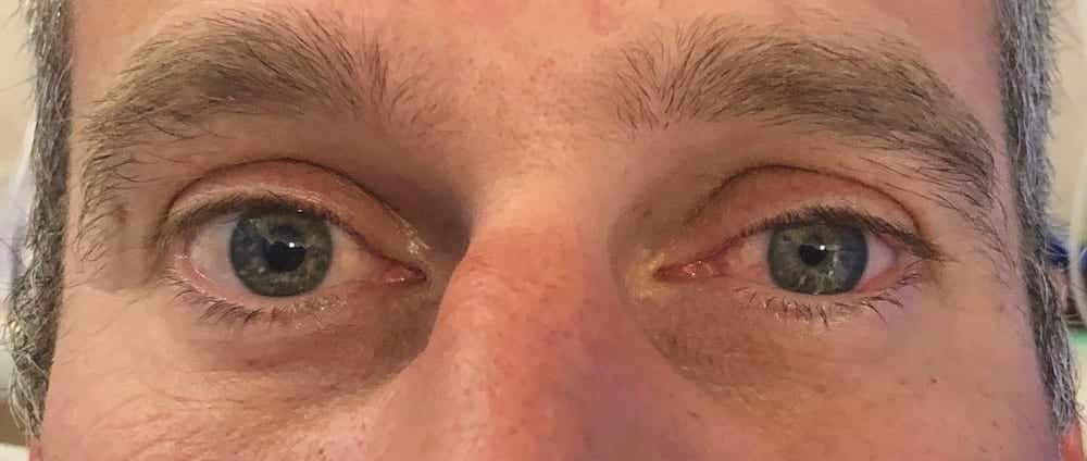
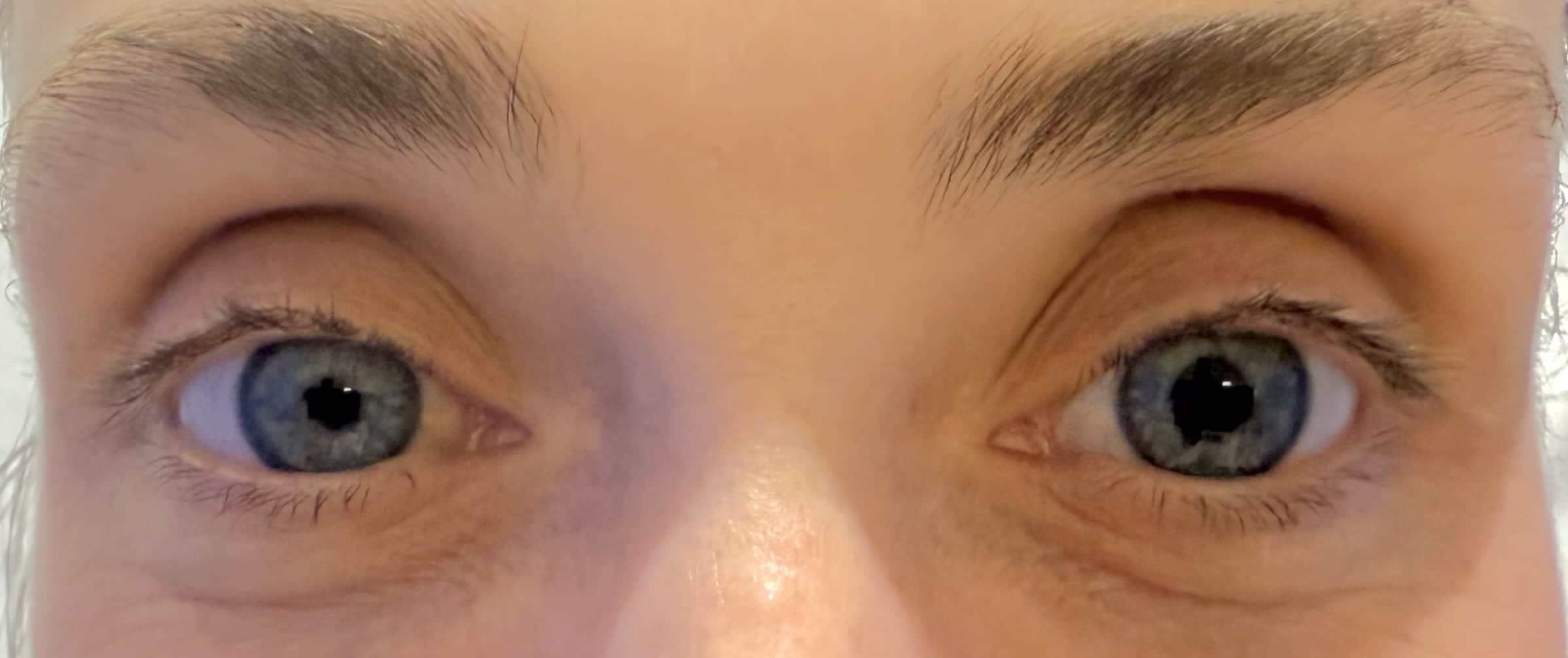
Horner syndrome, also known as oculosympathetic paresis, is a neurological disorder caused by disruption of the sympathetic pathway from the hypothalamus to the eye and face. It manifests with a classic triad of
- Ptosis: Drooping of the upper eyelid due to paralysis of the superior tarsal muscle
- Miosis: Constricted pupil due to loss of sympathetic innervation to the dilator pupillae muscle
- Anhidrosis: Absence of sweating on the affected side of the face
Horner first described the condition clinically in 1869, expanding on earlier experimental work by Claude Bernard (1813-1878). His description helped localize the lesion within the sympathetic chain and distinguish central from peripheral lesions.
1869 – Horner reported the findings of ptosis, miosis, enophthalmos in a 40-year-old peasant woman in Über eine Form von Ptosis. He also observed increased skin temperature and dryness of the ipsilateral face.
He pharmacologically confirmed the impairment of sympathetic innervation to the eye after noting poor dilation of the affected pupil following instillation of atropine and preserved pupillary constriction to the parasympathomimetic agent calabar. [1869;7:193-198]
Original
English
Frau Anna Brändli, 40 Jahre alt, eine gesund aussehende Bäuerin mittlerer Grösse… bemerkte ein allmähliges herabsinken des rechten oberen Augenlides, das sehr langsam zunahm… Das obere Lid deckt die rechte Cornea bis an den oberen Pupillarrand… Die Pupille des rechten Auges ist bedutend enger als diejenige des linken… Nun erst erzählte uns die Patientin, dass sie rechterseits nie geschwitzt habe
Die Untersuchungen ergaben also Integrität des sensiblen Trigeminus, transitorische Lähmung des vasomotorischen Fasern im rechtseitigen Trigeminusgebiet;… (es) wundert sich Niemand, wenn ich diese… nie vollständige Ptosis als Lähmung des vom Sympathicus versorgten organischen Musc. palpebral super. ansehe.
Anna Brändli, aged 40, healthy looking peasant woman…noticed a slight drooping of her right upper eyelid, which increased very gradually… The upper lid covers the right cornea to the upper edge of the pupil; the lid is not loose or wrinkled but somewhat sunken into the orbit and is still capable of movement; it is neither injected nor swollen.
The pupil of the right eye is considerably more constricted than that of the left, but reacts to light; the globe has sunk inward very slightly. During the clinical discussion … the right side of her face became red and warm; while the left side remained pale and cool.
The vasomotor disturbance involves not only the trigeminal area, but also the fibres of the cervical sympathetic; this experiment with belladonna and calabar speaks for the dual control of the movements of the iris in man… we are dealing with right dilator paralysis. Ptosis a paralysis of the musculus palpebrae superioris supplied by the sympathetic nerve (H. Muller, Harling), and the appearance of the upper lid as part and parcel of the whole symptom-complex.
Müller’s muscle (Horner’s muscle)
Circular fibres that are the innermost portion of the ciliary muscle; the lacrimal portion of the orbicularis oculi muscle (tensor tarsi muscle). Named after Heinrich Müller (1820-1864), but sometimes referred to as Horner’s muscle as it is affected in Horner syndrome.
Major Publications
- Horner JF. Über die Krümmung der Wirbelsäule im Aufrechten Stehen. Thesis 1854
- Horner F. Über eine Form von Ptosis. Klinische Monatsblätter für Augenheilkunde 1869;7:193-198 [Horner syndrome]
- Horner F. On spectacles: their history and uses. 1887. The Society for the Prevention of Blindness.
- Horner Fr. Die Krankheiten des Auges im Kindesalter. (The diseases of the eye in childhood) 1882.
- Horner Fr. Beiträge zur Ophthalmologie als Festgabe Friedrich Horner: zur Feier des fünfundzwanzigjährigen Jubiläums seiner academischen Lehrthätigkeit gewidmet. (Contributions to ophthalmology Friedrich Horner:25 year anniversary) 1881
- Horner JF. Ein Lebensbild geschrieben von ihm selbst. (Friedrich Horner autobiography) 1887.
References
Biography
- Johann Friedrich Horner, (1831-1886) “a form of ptosis”. JAMA. 1969 Jun 9;208(10):1899-900.
- Thompson HS. Johann Friedrich Horner (1831-1886). Am J Ophthalmol. 1986 Dec 15;102(6):792-5.
- Van der Wiel HL. Johann Friedrich Horner (1831-1886). Journal of Neurology, 2002; 249(5), 636–637
- Roper-Hall G. Historical Vignette: Johann Friedrich Horner (1831-1886): Swiss Ophthalmologist, Scientific Contributor, and Accomplished Academician. Am Orthopt J. 2016 Jan;66(1):126-134.
- Fustes OJH, Kay CSK, Lorenzoni PJ, Ducci RD, Werneck LC, Scola RH. Horner syndrome: tribute to Professor Horner on his 190th birthday. Arq Neuropsiquiatr. 2021 Jul;79(7):647-649.
Eponymous terms
- Shinohara H, Kominami R, Yasutaka S, Taniguchi Y. The anatomy of the lacrimal portion of the orbicularis oculi muscle (tensor tarsi or Horner’s muscle). Okajimas Folia Anat Jpn. 2001 Mar;77(6):225-32
- Borges AF. The original Z-plasty. British Journal of Plastic Surgery. 26 (3): 237–246.
- Koczman J. Vernal Keratoconjunctivitis. uiowa Eye Rounds 2007.
- Abbas A, Manjila S, Singh M, Belle V, Chandar K, Miller JP. Johann Friedrich Horner and the Repeated Discovery of Oculosympathoparesis: Whose Syndrome Is It? Neurosurgery. 2015 Sep;77(3):486-91
- Dafereras M, Sapouridis H, Laios K, Chrysikos D, Mavrommatis E, Troupis T. The pioneer ophthalmologist Johann Friedrich Horner (1831-1886) and the clinical anatomy of the homonymous syndrome. Acta Chir Belg. 2020 Oct;120(5):363-365.
Eponym
the person behind the name
BA MA (Oxon) MBChB (Edin) FACEM FFSEM. Emergency physician, Sir Charles Gairdner Hospital. Passion for rugby; medical history; medical education; and asynchronous learning #FOAMed evangelist. Co-founder and CTO of Life in the Fast lane | On Call: Principles and Protocol 4e| Eponyms | Books |