


Köhler teardrop
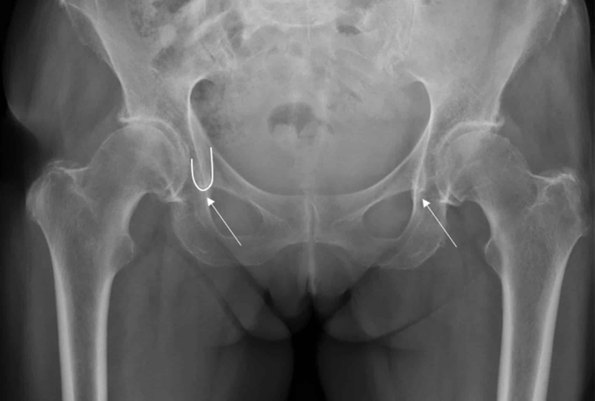
Köhler teardrop (Köhler tear; pelvic teardrop; Köhlersche Tränenfigur; teardrop; U-figure) is a U- or teardrop-shaped radiographic summation shadow seen medial to the acetabulum on an AP pelvis radiograph. It lies between the inferomedial acetabular region and the ilioischial (Köhler) line and does not correspond to a single anatomical structure.
In a normally positioned pelvis, the teardrop is formed by tangential projection of several bony surfaces. Anatomical studies identify the:
- the lateral limb with cortex in the anterior part of the acetabular fossa,
- the medial limb with the medial pelvic wall near the plane of the posterior acetabular margin, and
- the inferior arc with the superior anterior margin of the obturator foramen.
Because different cortical surfaces become tangential to the X-ray beam with pelvic rotation and tilt, the shape and position of the teardrop vary with projection.
The teardrop is used as a landmark in assessment of acetabular morphology and dysplasia, acetabular fractures, protrusio acetabuli, hip-joint effusion and postoperative hip arthroplasty measurements. Its use as an absolute landmark for reconstructing the centre of rotation after total hip arthroplasty is limited by patient-related variation, although it remains useful for serial intraindividual comparison.
Köhler teardrop and Köhler line are related but distinct radiographic landmarks. The teardrop is a composite U-shaped summation shadow produced by several acetabular and pelvic cortical surfaces. The Köhler line is the longer ilioischial contour representing the projected posterior column. The medial limb of the teardrop shares part of the projectional anatomy of Köhler line, but the terms are not synonymous. See also: Köhler line.
History of the Köhler teardrop
1905 – Alban Köhler (1874–1947) described the teardrop-shaped radiographic appearance in Die normale und pathologische Anatomie des Hüftgelenks und Oberschenkels in röntgenographischer Darstellung. Medial to the large acetabular contour he identified an elongated, superiorly open curved figure “…etwa von der Form eines herabfallenden Wassertropfens oder einer sogenannten Glasthräne.” […approximately in the form of a falling water drop or a so-called glass tear.]
Köhler investigated the appearance using lead-wire markers, skeletal preparations, stereoscopic radiographs and multiple projections. He found that the lines did not correspond simply to visible bony ridges or edges, but arose from projected surfaces within and around the acetabulum. He observed that the configuration changed with age and X-ray projection. He found it was relatively consistent in children but more dependent on tube position and pelvic orientation in adults.
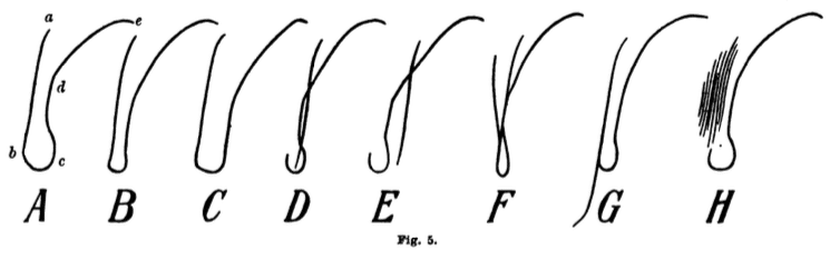
1910 — In the first edition of Lexikon der Grenzen des Normalen und der Anfänge des Pathologischen im Röntgenbilde, Köhler incorporated the appearance into his general radiology reference work. He wrote that the three projected lines formed a figure remotely resembling a tear, which he had therefore called the Tränenfigur, adding that the term had already found general acceptance. The four principal projectional configurations were illustrated as Fig. 49 A–D.
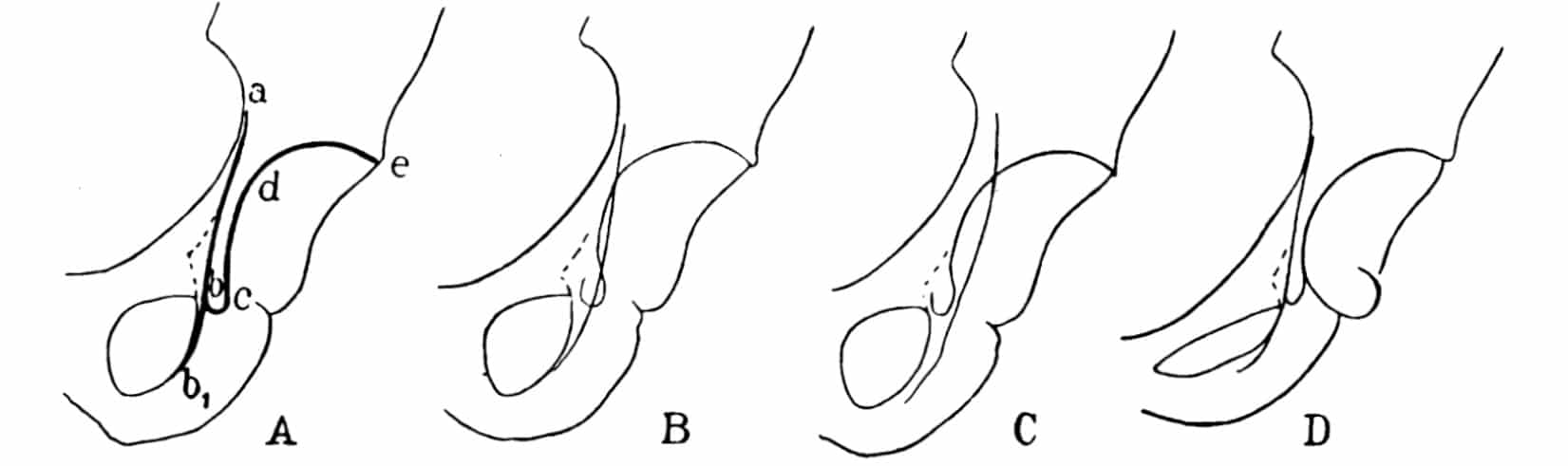
1952 – Victor B. Vare Jr. experimentally investigated the anatomical basis of the pelvic tear figure. Using lead foil applied around the acetabular fossa and extending onto the medial pelvic wall, he demonstrated the projected course contributing to the teardrop on AP radiographs.
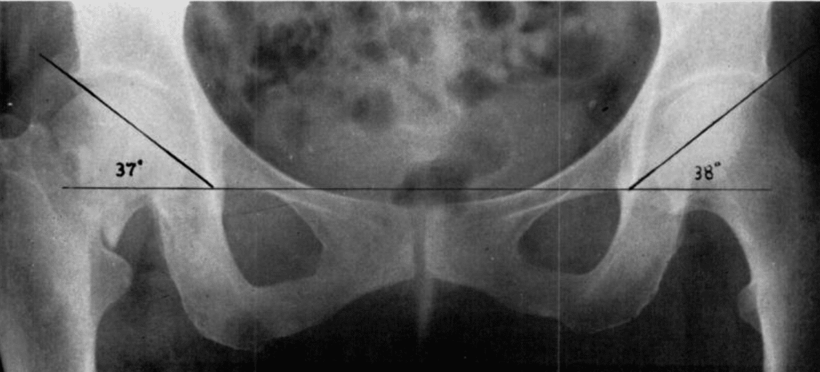
1961 – Ian K. Sharp used the inferior tip of the U-figure or “pelvic tear drop” as the medial landmark for his acetabular angle. Sharp defined the teardrop as a projectional structure and investigated the effect of pelvic tilt on the measurement. In 200 adult hips, normal acetabular angles were predominantly 33–38°.
1964 – Robert Judet, Jean Judet and Émile Letournel incorporated the “roentgenographic U” into their systematic radiographic assessment of acetabular fractures. They described its lateral limb as arising from the inferoanterior acetabular fossa and its medial limb from the anterior quadrilateral surface, noting that the U normally intersects or lies tangent to the ilioischial line. Displacement or separation of these landmarks assisted localisation of acetabular column fractures.
1977 – R. Kölbel and H. Golzo undertook a detailed experimental study of the anatomical substrate of the Köhlersche Tränenfigur. They used serial radiographs with progressive pelvic rotation and tilt, and radiographs of serial bone sections. They demonstrated that the teardrop was a dynamic summation shadow, with different cortical surfaces contributing according to projection. In the normal AP position they identified:
- lateral limb: a narrow 2 mm cortical region in the anterior acetabular fossa, around the transition from the middle to anterior third;
- inferior arc: the horizontal superior-anterior border of the obturator foramen;
- medial limb: the medial pelvic wall approximately in the plane of the posterior acetabular margin.
Original
English
Durch unsere Untersuchungen konnte belegt werden, daß die Kortikalisbereiche … bei definierten Beckendrehungen einem dynamischen Lagewechsel unterliegen bzw. daß jeweils andere Kortikalisanteile abgebildet werden.
Our investigations demonstrated that the cortical regions contributing to the teardrop undergo a dynamic change with defined pelvic rotation. Different cortical areas are therefore projected in different positions.
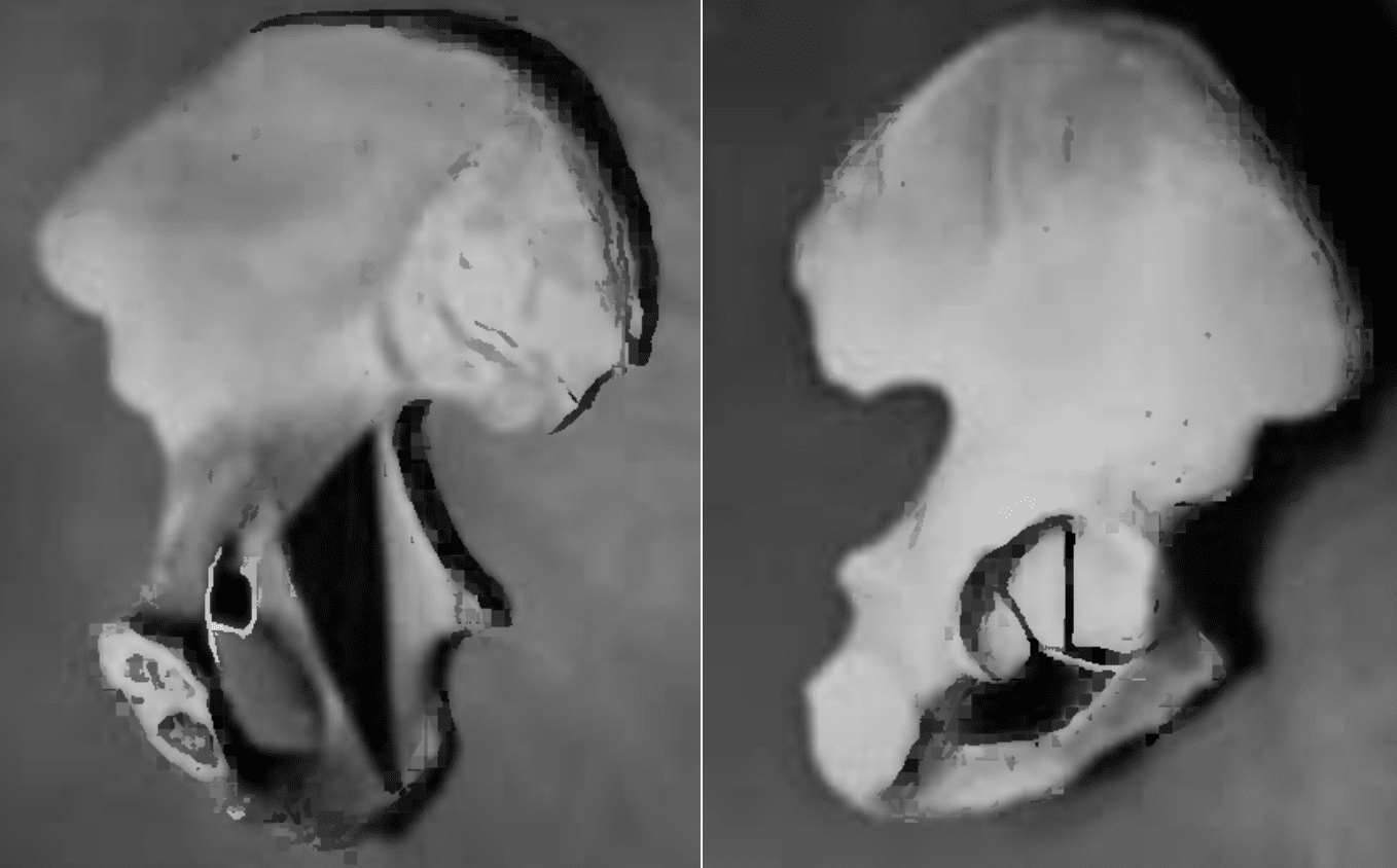
1982 – Jack W. Bowerman, Joseph M. Sena and Richard Chang published The teardrop shadow of the pelvis; anatomy and clinical significance. removing sections of the innominate bone to identify the structure responsible for the teardrop shadow. They localised it to cortical struts in the anteroinferior acetabular fossa at the acetabular notch, mainly from the ischium with a smaller pubic contribution. Removal abolished the teardrop and replacement restored it. They also demonstrated that loss or deformity of the teardrop could indicate destructive acetabular disease.
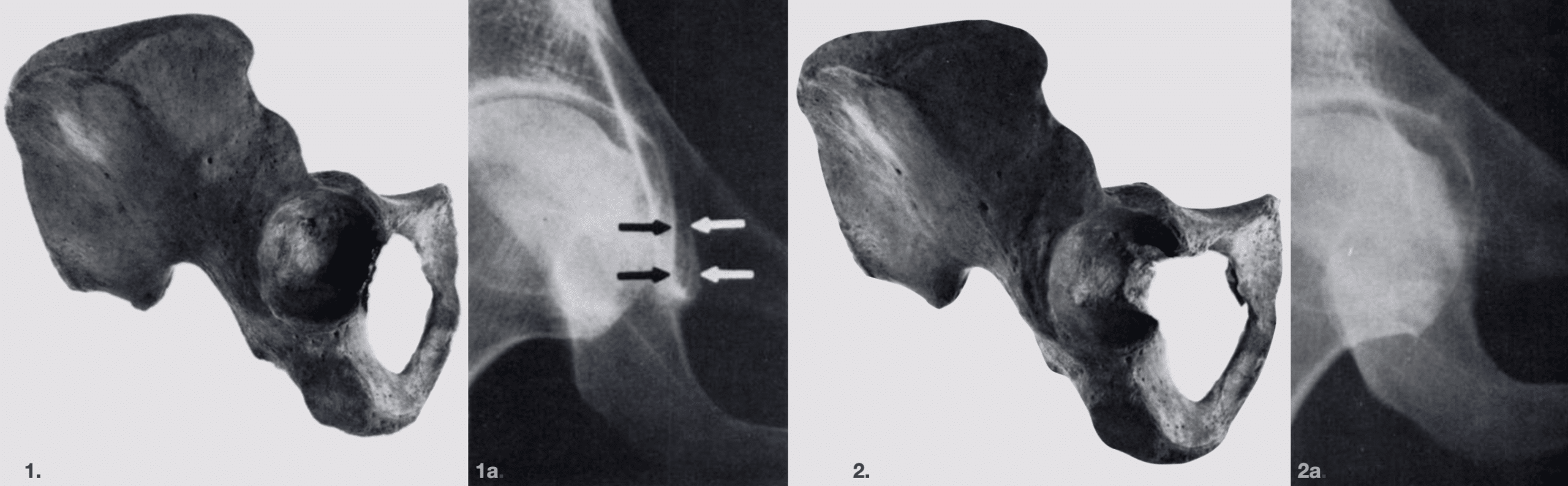
Bowerman et al., 1982. 1. Right innominate bone viewed from the lateral aspect. The darkened area is responsible for the teardrop and represents the inferior aspect of the anterior part of the superior ischiopubic junction. 1a. Anteroposterior radiograph of the intact innominate bone; the arrows outline the teardrop shadow.
2. The same innominate bone after removal of the area responsible for the teardrop. 2a. Anteroposterior radiograph after resection, showing that the teardrop shadow is no longer present.
1987 – John P. Sweeney et al. defined the teardrop distance as the distance between the lateral margin of the pelvic teardrop and the medial femoral head. In their small adult series, side-to-side widening of ≥1 mm was associated with hip effusion, with reported sensitivity of 90% and specificity of 100% in the absence of degenerative disease. They cautioned that the superimposed ilioischial line should not be confused with the medial boundary of the measurement.
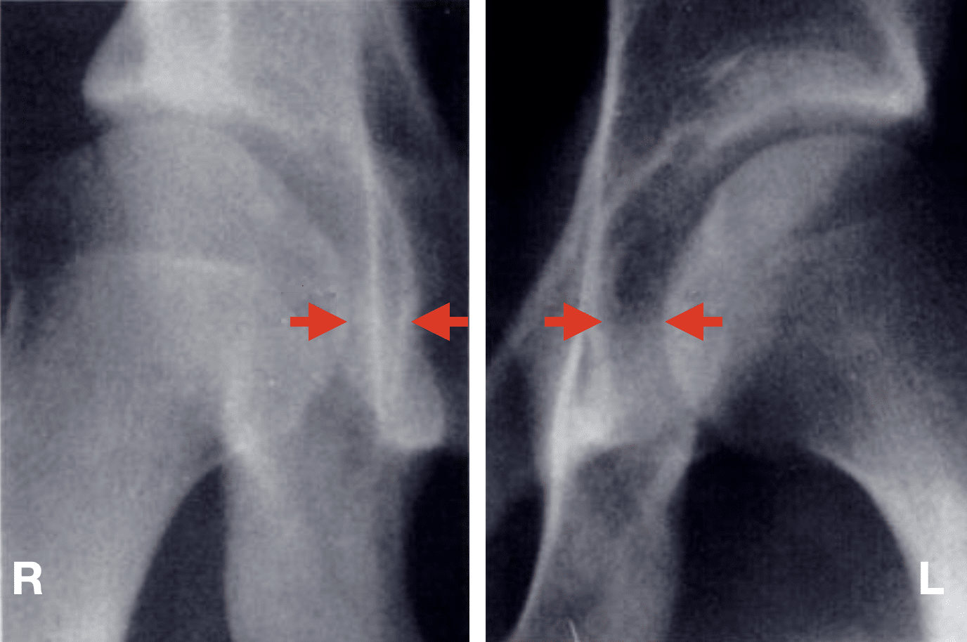
2023 – Heinz et al. evaluated the Köhler teardrop as a landmark for the centre of rotation after total hip arthroplasty. In 250 pelvic radiographs, its relationship to the hip centre varied with sex, age, height and weight. Pelvic tilt had relatively little effect, but the authors concluded that the teardrop was not sufficiently reliable as an absolute landmark for centre-of-rotation reconstruction, while remaining useful for intraindividual serial comparison.
Associated Persons
- Alban Köhler (1874–1947)
Eponymythology
Köhler’s attribution is supported by his 1905 primary description. He compared the appearance with a Wassertropfen (“water drop”) and Glasthräne (“glass tear”), and later Tränenfigur (“tear figure”). In German literature the term Köhlersche Tränenfigur was used and in English-language literature the terms teardrop, pelvic teardrop or U-figure.
The Köhler teardrop should not be confused with the Köhler line. Both emerged from Köhler’s investigation of the radiographic anatomy of the acetabulum and medial pelvic wall, but they describe distinct projected landmarks.
References
Historical references
- Köhler A. Die normale und pathologische Anatomie des Hüftgelenks und Oberschenkels in röntgenographischer Darstellung. Hamburg: Lucas Gräfe & Sillem; 1905: 15-24
- Köhler A. Lexikon der Grenzen des normalen und der Anfänge des pathologischen im Röntgenbilde. 1910: 95-99
- Köhler A. Grenzen des normalen und Anfänge des pathologischen im Röntgenbilde. 4e, 1924: 160-164
- Köhler A. Röntgenology: The Borderlands of the Normal and Early Pathological in the Skiagram. Trans. Arthur Turnbull. William Wood. 1928: 187-194
- Hubbard MJ. The measurement of progression in protrusio acetabuli. Am J Roentgenol Radium Ther Nucl Med. 1969 Jul;106(3):506-8.
Eponymous term review
- Vare VB Jr. The anatomy of the pelvic tear figure. J Bone Joint Surg Am. 1952 Jan;34-A(1):167-9.
- Sharp IK. Acetabular dysplasia: the acetabular angle. J Bone Joint Surg Br. 1961;43-B(2):268–272.
- Judet R, Judet J, Letournel E. Fractures of the acetabulum: classification and surgical approaches for open reduction. Preliminary report. J Bone Joint Surg Am. 1964 Dec;46:1615-46.
- Kölbel R, Golzo H. Die Köhlersche Tränenfigur. Untersuchungen zur Beziehung von Röntgenbild und anatomischem Substrat [Köhler’s “tear drop”. Relationship between the radiologic picture and anatomic substrate]. Rofo. 1977 Oct;127(4):326-33.
- Bowerman JW, Sena JM, Chang R. The teardrop shadow of the pelvis; anatomy and clinical significance. Radiology. 1982 Jun;143(3):659-62.
- Sweeney JP, Helms CA, Minagi H, Louie KW. The widened teardrop distance: a plain film indicator of hip joint effusion in adults. AJR Am J Roentgenol. 1987 Jul;149(1):117-9
- Heinz K, Nowack D, von Eisenhart-Rothe R, Wassilew G, Matziolis G, Brodt S. “Koehlers teardrop is not a reliable landmark for assessing the centre of rotation after Total hip arthroplasty” – a retrospective radiological study. Arch Orthop Trauma Surg. 2023 Sep;143(9):5671-5676.
- Kyle E, Cadogan M. Hip and pelvic lines and arcs. LITFL
eponymictionary
the names behind the name

BA MA (Oxon) MBChB (Edin) FACEM FFSEM. Emergency physician, Sir Charles Gairdner Hospital. Passion for rugby, medical history, medical education, and asynchronous learning #FOAMed evangelist. Editor-in-Chief and Webmaster of Life in the Fast lane, LITFL | On Call 4e| Eponyms | Books | Horology |