


Köhler line
Köhler line (ilioischial line) is a radiographic landmark on the anteroposterior pelvis used to assess the medial relationship of the acetabulum and femoral head to the pelvis.
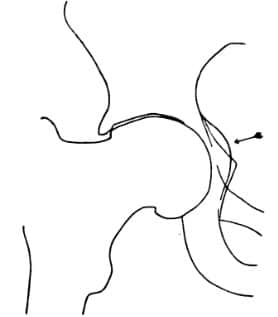
The line follows the medial pelvic contour from the ilium, passing tangentially along the medial acetabular region to the medial border of the ischium. In the normal hip, both the medial acetabular wall and femoral head remain lateral to the ilioischial line.
It can be used to distinguish coxa profunda from protrusio acetabuli:
- Normal: the medial acetabular wall and femoral head lie lateral to Köhler line.
- Coxa profunda: the medial acetabular wall projects medial to the line, while the femoral head remains lateral.
- Protrusio acetabuli: the femoral head itself projects medial to Köhler line.
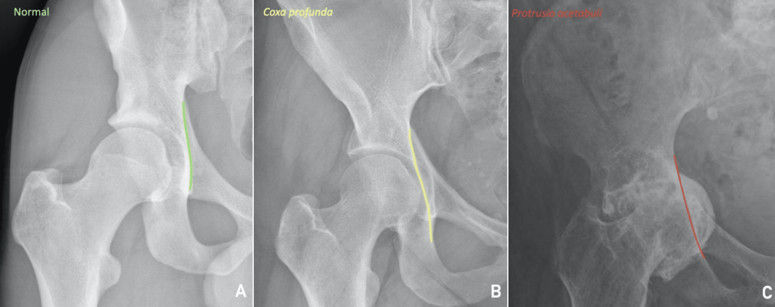
The Köhler line provides a simple reference for medial acetabular depth and femoral-head migration. An acetabular floor crossing the line represents a deep acetabulum, whereas extension of the femoral head medial to the line defines radiographic protrusio acetabuli.
Not to be confused with the Köhler teardrop, a composite U-shaped shadow adjacent to the medial acetabulum that Köhler investigated during the same studies of pelvic projectional anatomy.
History of the Köhler line
1905 – Alban Köhler (1874–1947) published Die normale und pathologische Anatomie des Hüftgelenks und Oberschenkels in röntgenographischer Darstellung. In his analysis of the normal acetabulum, Köhler investigated the multiple lines produced by projection of the acetabulum and pelvic wall using skeletal radiographs, stereoscopic images, sections and cadaver studies.
He labelled the long straight component a–b of his Thränenzeichnung (“tear figure”) and identified it as the tangential radiographic projection of the wall of the true pelvis anterior to the ischial spine, rather than a discrete anatomical edge.
Original
English
Der gerade lange Strich ab der Thränenzeichnung … entspricht also der Wandstelle des kleinen Beckens, welche nahe vor der Spina ischiadica sich flachzylindrisch gekrümmt von der inneren Tuberischiikante aus nach der tiefsten Einbuchtung der Incisura ischiadica major hinzieht. Köhler 1905
The long straight line a–b of the tear figure corresponds to that part of the wall of the true pelvis which, close anterior to the ischial spine, curves in a shallow cylindrical fashion from the medial border of the ischial tuberosity towards the deepest part of the greater sciatic notch. Köhler 1905
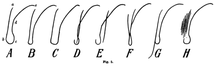
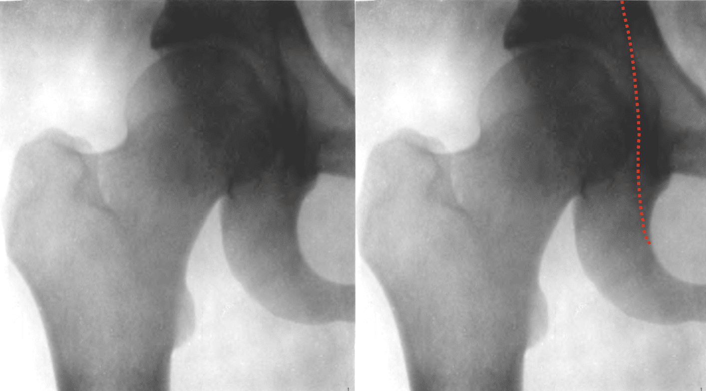
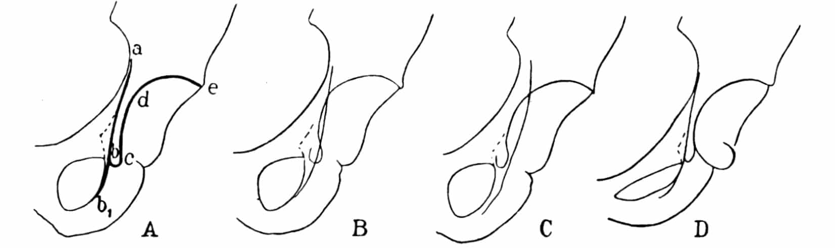
1910 – In the first edition of Lexikon der Grenzen des Normalen und der Anfänge des Pathologischen im Röntgenbilde, Köhler incorporated his hip studies into a general radiological reference work. He retained the three-line construction and illustrated four configurations produced by changes in X-ray projection. Köhler identified a–b–b₁ as the shadow of the wall of the true pelvis and emphasised that the apparent radiographic lines represented projected curved surfaces rather than anatomical edges.
Original
English
Die dritte Linie a b b₁ ist der Schatten der in der Strahlenrichtung verlaufenden Wand des kleinen Beckens…Diese drei Linien entsprechen also nicht irgendwelchen Kanten am Becken … sondern drei mehr oder weniger gekrümmten Flächen. Kohler 1910: 95
The third line a–b–b₁ is the shadow of the wall of the true pelvis running in the direction of the X-ray beam…These three lines therefore do not correspond to particular edges of the pelvis … but to three more or less curved surfaces. Kohler 1910: 95
1924 – In the fourth German edition, Köhler retained essentially the same description and four-view projection scheme. He also discussed traumatic displacement of the acetabular floor and illustrated protrusio acetabuli (Fig. 113), linking abnormal medial acetabular projection with intrapelvic protrusion.
1928 – Arthur Turnbull’s English translation of the fifth German edition appeared as Röntgenology: The Borderlands of the Normal and Early Pathological in the Skiagram. The established three-line analysis was retained, including Köhler’s statement “The third line a b b₁ is the shadow of the wall of the pelvis running in the direction of the rays“
The translation made Köhler’s acetabular projection anatomy readily accessible to English-speaking radiologists, while still not using the eponym “Köhler line.”
1939 – John Gilmour explicitly referred to “Kohler’s lines” in his study of protrusio acetabuli, based on his 1936 Hunterian Lecture. He reproduced Kohler’s 1910 figure and identified a–b–b₁ as the line produced by the inner wall of the pelvis and c–d–e as the acetabular contour.
Using a tracing of Fröhlich’s case, Gilmour demonstrated protrusio when the acetabular curve crossed medial to the pelvic-wall line “Kohler has described in detail the significance of the various ‘lines’ which characterize the region of the normal acetabulum on an X-ray film.”
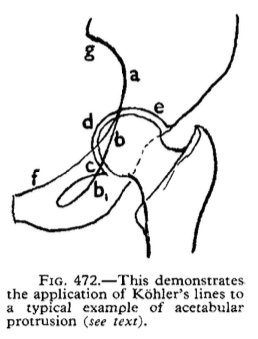
In Gilmour’s tracing of Fröhlich’s case, the acetabular line c–d–e crossed the pelvic-wall line a–b–b₁, while Shenton’s arc remained intact.
1969 – M. J. S. Hubbard used the singular term Köhler’s line and defined it in terms equivalent to the modern ilioischial line:
This line begins on the pelvic border of the ilium and ends on the medial border of the body of the ischium, brushing the dome of the acetabulum on its way.
Hubbard 1969
Hubbard used the perpendicular distance between the acetabular dome and Köhler’s line to quantify and serially monitor protrusio acetabuli. In normal hips the acetabular outline met or lay slightly lateral to the line; medial migration represented protrusion.
Hubbard measured the perpendicular distance between the acetabular dome and Köhler’s line to quantify serial progression of protrusio acetabuli. Hubbard cited Köhler’s 1935 second English edition, p.222 as his source.
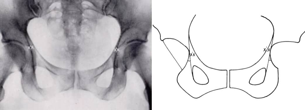
2005 – Modern radiographic descriptions generally use the anatomical term ilioischial line. Campbell described it on an AP pelvis radiograph as a line drawn from the ilium to the ischial tuberosity and identified it as part of the radiographic posterior column of the pelvis.
Associated Persons
- Alban Köhler (1874–1947)
Alternative names
- Kohler’s lines, Kohler’s line
- Köhler line
Eponymythology
Köhler did not introduce the term “Köhler line”. In 1905 he labelled its principal pelvic-wall component a–b and experimentally demonstrated that it represented the tangential projection of a curved portion of the true pelvic wall. In 1939, Gilmour referred collectively to “Köhler’s lines” and applied them to assessment of protrusio acetabuli. In 1969, Hubbard used the singular “Köhler’s line” for the ilioischial contour used today.
The eponym has evolved from Köhler’s original anatomical work. The radiographic concept is derived from Köhler’s investigations, and the eponymous name introduced by later authors.
References
Historical references
- Köhler A. Die normale und pathologische Anatomie des Hüftgelenks und Oberschenkels in röntgenographischer Darstellung. Hamburg: Lucas Gräfe & Sillem; 1905: 15-24
- Köhler A. Lexikon der Grenzen des normalen und der Anfänge des pathologischen im Röntgenbilde. 1910: 95-99
- Köhler A. Grenzen des normalen und Anfänge des pathologischen im Röntgenbilde. 4e, 1924: 160-164
- Köhler A. Röntgenology: The Borderlands of the Normal and Early Pathological in the Skiagram. Trans. Arthur Turnbull. William Wood. 1928: 187-194
- Frohlich M. Migration intrapelvienne progressive da la tête fémorale et lésion de l’ovaire. Revue d’orthopédie et de chirurgie de l’appareil moteur 1930; 17: 553.
- Gilmour J. Adolescent deformities of the acetabulum an investigation into the nature of protrusio acetabuli, British Journal of Surgery, 1939; 26(104): 670–699
- Hubbard MJ. The measurement of progression in protrusio acetabuli. Am J Roentgenol Radium Ther Nucl Med. 1969 Jul;106(3):506-8.
Eponymous term review
- Campbell SE. Radiography of the hip: lines, signs, and patterns of disease. Semin Roentgenol. 2005 Jul;40(3):290-319.
- Bi AS, Carter C, Price AE, Litrenta J, Karamitopoulos M, Castañeda PG. A history of eponym usage in hip and pelvis radiography part 1: the paediatric hip. Hip Int. 2023 Mar;33(2):136-143.
- Bi AS, Ryan MK, Castañeda PG, Youm T. A history of eponym usage in hip and pelvis radiography part 2: the adult hip. Hip Int. 2023 Mar;33(2):144-151
- Kyle E, Cadogan M. Hip and pelvic lines and arcs. LITFL
eponymictionary
the names behind the name

MB BAO BCh Trinity College Dublin, MSc Clinical Data Analytics University of Galway. Junior doctor in Perth, doctoral researcher in MRI radiomics. Working towards radiology training.

BA MA (Oxon) MBChB (Edin) FACEM FFSEM. Emergency physician, Sir Charles Gairdner Hospital. Passion for rugby, medical history, medical education, and asynchronous learning #FOAMed evangelist. Editor-in-Chief and Webmaster of Life in the Fast lane, LITFL | On Call 4e| Eponyms | Books | Horology |