![]()

Echo basics: Aortic Valve
Echo basics: Aortic Valve. A normal aortic valve is trileaflet, with equally sized cusps that are supported by a fibrous annulus and separated by three commissures.

![]()
Echo basics: Aortic Valve. A normal aortic valve is trileaflet, with equally sized cusps that are supported by a fibrous annulus and separated by three commissures.
Echocardiography basics. Grading and quantifying mitral stenosis (MS) with planimetry, pulsed wave Doppler, PHT and Continuity Equation Method
Mitral regurgitation (MR) is a common pathology detected during echocardiography. Accurate identification and grading rely heavily on colour and spectral Doppler imaging across multiple standard views.
The mitral valve is a dominant structure in most standard echocardiographic views. Understanding its anatomy in each window is essential for accurate assessment.

McGinn and White first described the so-called S1Q3T3 pattern in five patients with acute cor pulmonale secondary to pulmonary embolism.
Echocardiography and valve measurements. Comprehensive assessment requires measurements to be made from 2D images and the waveforms generated during Doppler investigations
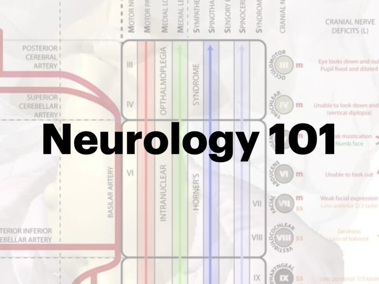
Neuro 101: Neurological Examination. The eight steps, mental status, motor, sensory, reflex, cerebellar examinations
Echocardiography and valve views. Overview of valve disease and parasternal, apical and subcostal valve views with the echo probe
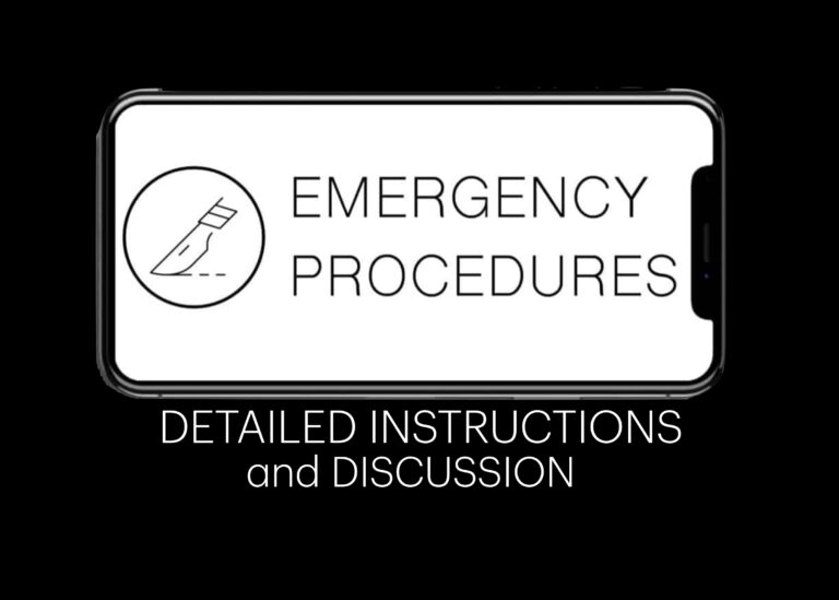
Emergency procedure, instructions and discussion: Tracheostomy emergencies for patients in respiratory distress or following accidental decannulation
Emergency procedure, instructions and discussion: Needle Cricothyroidotomy when unable to oxygenate and ventilate with (BVM, LMA, ETT) and age <10years
Emergency procedure, instructions and discussion: Laryngeal mask airway (LMA) for airway compromise and deeply reduced level of consciousness or arrest
Emergency procedure, instructions and discussion: Oropharyngeal airway for upper airway obstruction (partial or complete) and reduced level of consciousness