Ultrasound Case 038
Presentation
A 46 year old woman presents with relatively sudden painless visual field loss. She describes preceding flashers and floaters, and then a shadow falling over the medial and central part of her visual field.
Describe and interpret these scans
IMAGE INTERPRETATION
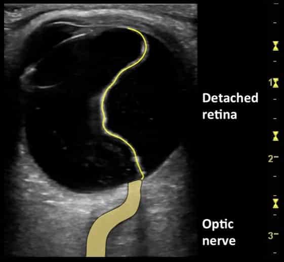
Image 1: Transverse image of left eye. The echogenic retinal flap is seen moving with occular movement. It attaches posteriorly at the optic nerve. Retinal fibers pass into the optic nerve and detachment cannot occur at this point.
There is an associated vitreous detachment – much finer swirling debris is seen in the posterior chamber.
Image 2: Transverse image of the left eye with label. (with annotation below)
CLINICAL CORRELATION
Retinal detachment
Vitreous detachment can also cause a swirling linear appearance in the posterior chamber.
Key differences between Vitreous and Retinal detachment:
- Retinal membrane far thicker and brighter than vitreous detachment
- Retinal detachment is anchored at the optic nerve and it flaps with eye movement rather than the vitreous which tends to swirl more freely.
- I do not use Doppler to assess the flap of a retinal detachment – although there should be vascularity in the retinal membrane and not in a vitreous detachment. Movement artifact complicates assessment.
[cite]
TOP 100 ULTRASOUND CASES
An Emergency physician based in Perth, Western Australia. Professionally my passion lies in integrating advanced diagnostic and procedural ultrasound into clinical assessment and management of the undifferentiated patient. Sharing hard fought knowledge with innovative educational techniques to ensure knowledge translation and dissemination is my goal. Family, wild coastlines, native forests, and tinkering in the shed fills the rest of my contented time. | SonoCPD | Ultrasound library | Top 100 | @thesonocave |


