![]()
Ultrasound Case 039
Presentation
A 62 year old male attended the Emergency Department after tripping on the bottom step whilst ascending a flight of stairs. He has bilateral knee pain and has been unable to walk since the incident.
View 2: Clinical Video
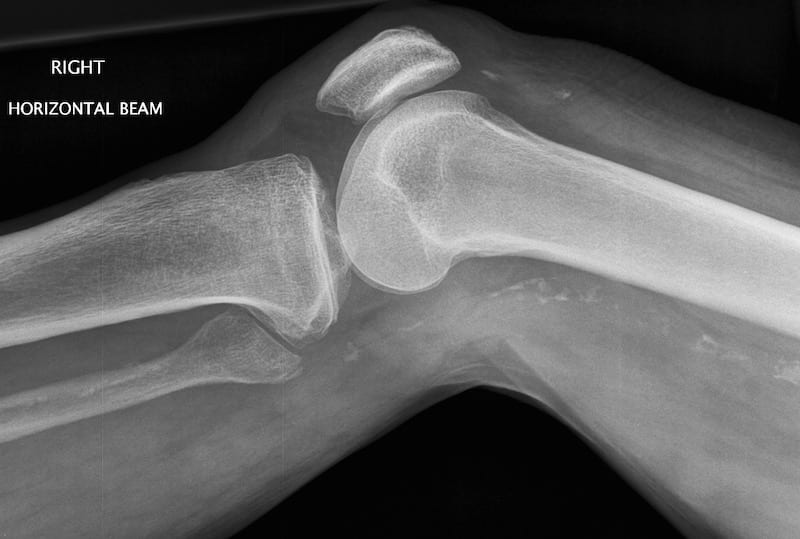
View 3: Knee X-Ray
View 4

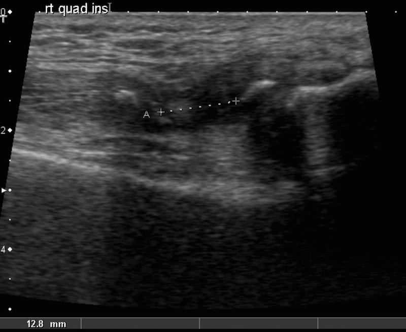
View 5
Describe and interpret these scans
IMAGE INTERPRETATION
Image 1: Ultrasound of his right quadriceps tendon at the site of insertion into the patella tendon, longitudinal section. Dynamic scan where he is asked to attempt a straight leg raise (SLR). The patella is seen on the right of the screen. As the SLR begins the patella and quadriceps tendon move as two distinct entities rather than sliding as one. The rupture with complete separation of the tendon from the patella is seen toward the end of the loop. The femur is seen posteriorly, and as the knee flexes the medial femoral condyle with its hypoechoic cartilaginous surface is seen.
Image 2: Clinical video; attempted SLR; identical appearance bilaterally.
Image 3: Plain film lateral knee; the avulsed quadriceps tendon is evident with small fragments of bone.
Image 4: Still image of the rupture; demonstrating why dynamic loops are far better at detecting and demonstrating ligamentous rupture.
Image 5: The defect between and two ends of the ruptured quadriceps is measured. A small avulsion fragment is seen in the proximal part.
CLINICAL CORRELATION
Quadriceps rupture
Injuries such as these are generally clinically apparent. When ultrasound is used to contribute to the assessment, dynamic scanning is important. Extending the musculotendinous complex will cause separation of the ends at the point of rupture, and usually a hypoechoic blood collection is seen between the two ends. Scanning in the neutral position and recording still images is less rewarding. The artifact known as anisotropy can be misinterpreted as a tear or rupture, and genuine tendinous disruptions can be missed as the ends are often apposed at rest.
TOP 100 ULTRASOUND CASES

An Emergency physician based in Perth, Western Australia. Professionally my passion lies in integrating advanced diagnostic and procedural ultrasound into clinical assessment and management of the undifferentiated patient. Sharing hard fought knowledge with innovative educational techniques to ensure knowledge translation and dissemination is my goal. Family, wild coastlines, native forests, and tinkering in the shed fills the rest of my contented time. | SonoCPD | Ultrasound library | Top 100 | @thesonocave |